


Page 89 - 南京医科大学自然版
P. 89
第45卷第2期          沈文婷,徐 怡,王云飞,等. 升主动脉局限性内膜撕裂CT血管成像的影像学特征分析[J].
                  2025年2月                     南京医科大学学报(自然科学版),2025,45(2):227-232                        ·229 ·


                                          表1 AD组、IMH组、LIT组与正常对照组临床资料比较
                          Table 1 Comparison of clinical data among AD group, IMH group,LIT group,and control group

                          Clinical data      AD group(n=120) IMH group(n=52) LIT group(n=40) Control group(n=212)  P
                Age(years,x ± s)                 54 ± 14        63 ± 10       59 ± 19        55 ± 14       0.002
                Male[n(%)]                      99(82.5)       30(57.7)      27(67.5) *     132(62.3)     <0.001
                Hypertension[n(%)]              88(73.3)       38(73.1)      30(75.0) #      73(34.4)     <0.001
                Hyperlipemia[n(%)]              12(10.0)        2(3.8)        5(12.5)        21(9.9)       0.486
                Diabetes[n(%)]                   6(5.0)         3(5.8)        2(5.0)         35(16.5)      0.003
                Smoking[n(%)]                   37(30.8)       11(21.2)      12(30.0)        43(20.3)      0.131
                Thoracic aortic atherosclerosis[n(%)]  54(45.0)  35(67.3)    26(65.0) *#     63(29.7)     <0.001
                                                                                            #
                                                                 *
                   P represents the difference among groups;compared with the AD group,P < 0.05;compared with the control group,P < 0.05.
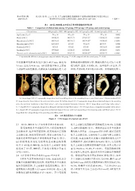
                主动脉瓣环的距离为 32.5(20.3,49.5)mm,最近为                   脉隆起或轻微轮廓异常,撕裂内膜片在CTA上可表
                7.0 mm,最远为 90.0 mm。LIT 的影像学特点主要是                  现为线性、星状、T形或L形。其中线性LIT最多,共
                主动脉壁局限性撕裂,内膜缺损基底部偏心性主动                            25例,星状8例,T形3例,L形4例。典型病例见图1。


                                     A                        B                       C                       D
                   A:Linear⁃shaped LIT,CT angiography images show focal linear filling defect of the ascending thoracic aorta(black arrow). Volume⁃rendered(VR)
                CT image shows the linear defect of the aortic root(white arrow). B:Stellate⁃shaped LIT,CT angiography images show undermined edges in the ascending
                aorta,the tear inner membrane is free(black arrow),and a few intramural hematoma formation. VR CT image shows ovoid bulge(white arrow).
                C :T⁃shaped LIT,CT angiography images show elongated undermined edge(black arrow). VR CT image shows a triangular tear pattern(white arrow).
                D:L⁃shaped LIT,CT angiography images show undermined edges of the tear,with focal outpouching of the remaining aortic wall(black arrow). VR CT
                image shows the L⁃shaped bulge of the ascending aorta(white arrow).
                                                   图1 LIT典型病例的CTA图像
                                              Figure 1  CTA images of a typical case of LIT


                2.3  AD组、IMH组与LIT组影像学特征参数比较                       处升主动脉长径预测LIT的准确性为94.4%,长径越
                    除无病变处升主动脉短径外,3 组间病变处升                         大,LIT风险越高(表3)。将临床因素及升主动脉参
                主动脉长径、短径和管腔面积,病变处相应层面降                            数均纳入多因素二元Logistic回归分析,提示无病变
                主动脉长径、短径和管腔面积,邻近无病变处升主                            处升主动脉长径、无病变处升主动脉与降主动脉长
                动脉长径和管腔面积差异均有统计学意义(表 2)。                          径比值、高血压病史以及胸主动脉粥样硬化是 LIT
                LIT 组病变处及邻近无病变处主动脉重构指数                            的预测因子,此时准确性为 96.8%(表 3);与无病变
                均大于 AD 组及 IMH 组,病变处升主动脉扩张程                        处升主动脉长径模型的准确性比较,差异无统计学
                度均高于 AD 组、IMH 组,差异均具有统计学意义                        意义(96.8% vs. 94.4%,P > 0.05)。考虑到临床诊断
               (P均 < 0.05)。                                       的方便性与快速性,建议采用升主动脉长径预测模

                2.4  LIT影像学特征参数的Logistic回归分析                      型。绘制单因素和多因素的 ROC 曲线(图 2),其中
                    LIT 组与正常对照组进行比较,高血压以及合                        升主动脉长径预测 LIT 的 AUC 为 0.948,最佳 cut⁃off
                并胸主动脉粥样硬化的比例、无病变处升主动脉长                            值为40.5 mm,此时的灵敏度和特异度分别为87.5%、
                短径及管腔面积、升主动脉扩张程度均增高。将上                            94.3%,准确性可以达到 81.8%。当长径<29.5 mm
                述参数纳入单因素二元Logistic回归分析,提示上述                       时,LIT 发生率为 0;当长径>72.5 mm 时,LIT 发生率
                参数均是 LIT 的预测因子(P < 0.05)。其中无病变                    为95%。

