


Page 95 - 南京医科大学自然版
P. 95
第46卷第7期       李   巧,方   蕊,黄佳鑫,等. 蛋白质精氨酸甲基转移酶1在乳腺癌组织中的表达及临床意义[J].
                  2026年7月                     南京医科大学学报(自然科学版),2026,46(7):1037-1045                      ·1041 ·


                  A                 P<0.01                        B                   P<0.01
                       300                                               300
                       250                                               250
                      Transcript per million  200                      Transcript per million  150
                                                                         200
                       150
                       100
                                                                         100
                        50                                               50
                        0                                                 0
                           Normal  Stage1  Stage2  Stage3  Stage4            Normal  N0     N1     N2     N3
                          (n=114)(n=183)(n=615)(n=247)(n=20)                (n=114)(n=516)(n=362)(n=120)(n=77)
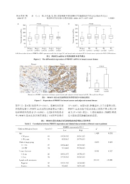
                A,B:Association between PRMT1 mRNA expression and clinical stage(A)and lymph node metastasis(B)in breast cancer patients in UALCAN database.
                                              图2 PRMT1 mRNA在乳腺癌组织中差异表达
                                 Figure 2 The differential expression of PRMT1 mRNA in breast cancer tissues


                               Normal                      Tumor
                                                                                      3         P<0.01


                                                                                     score  2

                                                                                     PRMT1  1


                                                                                      0
                                                                                           Normal   Tumor
                                          50 μm                       50 μm               (n=80)   (n=80)
                            Detection of PRMT1 expression in breast cancer tissues and adjacent normal tissues by IHC. The scale bar:50 μm.
                                         图3 PRMT1蛋白在乳腺癌组织和癌旁组织中的表达情况
                                  Figure 3 Expression of PRMT1 in breast cancer and adjacent normal tissues

                较早(Ⅰ~ⅡA期)的患者(P < 0.01)。在淋巴结转移                    (P < 0.05)。而在年龄、肿瘤直径、分子分型等方面,
                阳性的患者中,PRMT1高表达的比例显著高于淋巴                          PRMT1 高表达组与低表达组之间的差异无统计学
                结转移阴性的患者(P < 0.001)。在 ER 阴性的患者                    意义(P > 0.05,表 2)。上述结果提示,PRMT1 高表
                中,PRMT1 高表达的比例显著高于 ER 阳性的患者                       达可能促进乳腺癌的疾病进展。

                                        表2 PRMT1蛋白高表达与乳腺癌临床病理特征之间的关系
                         Table 2 Correlation between PRMT1 expression and clinical characteristics of breast cancer patients
                                                             PRMT1 expression[n(%)]
                  Clinicopathological feature  Cases(n)                                     χ 2          P
                                                             Low             High
                   Age                                                                     1.428        0.232
                     < 50 years                49          19(38.78)       30(61.22)
                     ≥ 50 years                31          08(25.81)       23(74.19)
                   Clinical stage group                                                    8.695        0.003
                     Ⅰ-ⅡA                      27          12(44.44)       15(55.56)
                     ⅡB-Ⅳ                      30          03(10.00)       27(90.00)
                   Tumor diameter                                                          0.961        0.327
                     ≤ 2 cm                    24          10(41.67)       14(58.33)
                     > 2 cm                    56          17(30.36)       39(69.64)
                   Lymph node metastasis                                                   14.110      < 0.001
                     Negative                  29          15(51.72)       14(48.28)
                     Positive                  34          03(8.82)        31(91.18)

