


Page 73 - 南京医科大学自然版
P. 73
第45卷第2期       陆  遥,邵文慧,宋佳成,等. 基于多参数磁共振的影像组学特征一致性聚类与早期宫颈癌临床及
                  2025年2月        组织病理学特征的相关性研究[J]. 南京医科大学学报(自然科学版),2025,45(2):208-217 ·213                   ·


                                                   A                     B                      C


                                                   D                     E                      F
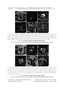
                   59⁃year⁃old woman in cluster 1 with preoperative stage IB2 cervical cancer and an MRI⁃detected tumor size of(25 mm×17 mm×17 mm)(long
                                                                                        2
                diameter×short diameter×height). A:Axial T2WI shows a regular lesion(arrow). B:On DWI(b value=1 000 s/mm ),the lesion shows high signal density
               (arrow). C,D:Lesion depicts decreased values on ADC map(C)and CE⁃T1WI(D). E:Sagittal view shows shallow cervical stromal invasion(arrow). F:
                Coronary fat⁃suppressed T2WI showes no apparent node. Postoperative pathological examination confirmed stage IB2 squamous cervical cancer with
                shallow(<1/3)stromal invasion,negative LNM and a tumor size of(27 mm×12 mm×5 mm)(long diameter × short diameter × height).
                                      图3 组1中的肿瘤具有较低临床及组织病理高危因素的典型病例
                      Figure 3 Typical case of a tumor in cluster 1 with lower risk clinical and histopathological high⁃risk factors


                                                   A                     B                      C


                                                   D                     E                      F
                   56⁃year⁃old woman in cluster 2 with preoperative stage IB3 cervical cancer and an MRI⁃detected tumor size of(48 mm×23 mm×25 mm)(long di⁃
                                                                                           2
                ameter × short diameter × height). A:Axial T2WI shows an irregular lesion(arrow). B:On DWI(b value=1 000 s/mm ),the lesion shows a high signal
                density(arrow). C,D:Lesion depicts decreased values on ADC map(C)and CE⁃T1WI(D). E:Sagittal view shows deep cervical stromal invasion(ar⁃
                row). F:Coronary fat⁃suppressed T2WI shows the left pelvic lymph node with the short axis of 5.5 mm(arrow). Postoperative pathological examination
                confirmes stage ⅢC1p squamous cervical cancer with deep(>2/3)stromal invasion,pelvic LNM and a tumor size of(46 mm×35 mm×16 mm)(long di⁃
                ameter × short diameter × height).
                                      图4   组2中的肿瘤具有较高临床及组织病理高危因素的典型病例
                      Figure 4 Typical case of a tumor in cluster 2 with higher risk clinical and histopathological high⁃risk factors

                更大的肿瘤直径。基于盆腔淋巴结的无监督聚类                                 放射组学作为一种非侵入性方法已经逐步应
                分析没有发现两组之间的差异性。                                   用于宫颈癌组织病理学预后因素、治疗反应、肿瘤

