


Page 162 - 南京医科大学自然版
P. 162
第45卷第7期
               ·1060 ·                           南 京    医 科 大 学 学         报                        2025年7月


                                               A                          B                         C
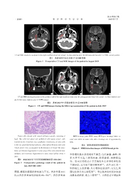
               CT and MRI revealed a neoplasm in the right maxillary sinus(red arrows). A:Bone window axial. B:Soft tissue window axial. C:T2WI coronal position.
                                              图1 患者2019年8月术前CT及MR影像
                                  Figure 1 Preoperative CT and MRI images of the patient in August 2019


                                               A                          B                         C
                 CT and MRI showing neoplasm in the posterior wall of the right maxillary sinus near the pterygopalatine fossa(red arrows). A:Bone window coro⁃
              nal. B:Soft tissue window axial. C:T2WI coronal.
                                            图2 患者2023年7月复诊期间CT及MR影像
                           Figure 2 CT and MRI images during the follow⁃up examination of the patient in July 2023


                 Tumor cells showed solid nested infiltrative growth,consisting of  MYB breakage probe FISH assay,MYB gene breakage(white ar⁃
              basal ⁃ like cells(red arrow)and epithelioid cells(green arrow),with  rows)was visible in tumor cells,with a breakage rate of approximately
              rounded nuclei of medium size,cytoplasmic translucency of some cells  60%.
                                                                           图4
              in the non⁃glandular luminal surfaces,a few nuclear divisions were seen  MYB断裂探针FISH检测
             (black arrow),foci accompanied by the formation of ductal⁃like struc⁃  Figure 4 FISH detection images of MYB break probe
              tures,and vitreous degeneration in some areas of the extra⁃tumoral mes⁃
              enchyme,and mucinous degeneration in some areas(yellow arrow,×
                                                                性肿瘤的临床表现通常不典型,包括鼻塞、鼻衄,体
              200).                                             积大者可引起上颌部疼痛、面部膨隆、硬腭隆起
                 图3   患者2023年7月术后病理检查结果(HE×200)
              Figure 3  Postoperative pathology result of the patient in  等。而 ACC 的特点在于其惰性生长和非特异性的
                                                                早期症状,这导致早期诊断困难 。此外 ACC 是一
                                                                                             [3]
                       July 2023(HE ×200)
                                                                种持续生长的肿瘤,具有嗜神经侵袭性,易发生周
                                                                                     [1]
              筛窦,蝶窦和额窦的肿瘤极为罕见。鼻腔鼻窦ACC                           围侵犯和多次局部复发 。常出现神经症状如颌面
                                             [1]
              约占鼻腔鼻窦恶性肿瘤的 4%~5% 。鼻腔鼻窦恶                          部麻木或疼痛,视力下降等            [4-5] 。本例患者早期因鼻

