Page 18 - 南京医科大学学报自然科学版
P. 18

第41卷第12期
               ·1724 ·                           南 京    医 科 大 学 学         报                        2021年12月


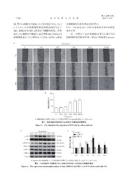
              制,用 C5a 刺激 PC9 细胞,在不同时间点(0 h、1 h、2                达和磷酸化均未见明显变化(图4)。
              h、3 h、6 h、12 h)检测增殖和迁移相关的信号分子                     2.2.4  C5a 通过 Akt1⁃ERK1/2 通路诱导 PC9 细胞增
              Akt1、ERK1/2和PKC⁃α的表达与磷酸化情况。结果                     殖和迁移
              显示,C5a刺激PC9细胞后,显著增强Akt1和ERK1/2                         进一步探讨了 Akt1 和 ERK1/2 在 C5a 诱导 PC9
              的磷酸化水平,且其峰值在3 h左右,而PKC⁃α的表                        细胞增殖和迁移的作用。用 Akt1 抑制剂 Perifosine

              A                                             C5a(ng/mL)

                        0              0.05            0.50           5.00           50.00          500.00


                 0 h


                 h
                 24


                 h
                 48


                                            B
                                                 150
                                                ( % )               *   *    *
                                                细胞迁移率 100


                                                  50


                                                   0
                                                      0   0.05 0.50  5.00 50.00 500.00
                                                              C5a(ng/mL)
                                   A:细胞划痕代表图片(×40);B:半定量柱形图(与0 ng/mL组比较,P < 0.01,n=3)。
                                                                               *
                                         图3 细胞划痕实验检测C5a刺激PC9细胞的迁移情况
                                   Figure 3 C5a stimulates the migration of PC9 cells by cell scratch test


                    A                 C5a(ng/mL)                 B
                                                                     4
                                                                         p⁃Akt1
                              0 h  1 h  2 h  3 h  6 h 12 h
                                                                         p⁃ERK1/2
                         p⁃Akt1                       —60 kDa        3   p⁃PKC⁃α         **
                                                                     蛋白相对表达量  2    *  *
                         t⁃Akt1
                                                      —60 kDa                           **
                       p⁃ERK1/2                       —42、44 kDa             *                 *     *
                       t⁃ERK1/2                       —42、44 kDa
                       p⁃PKC⁃α                                       1
                                                      —80 kDa
                        t⁃PKC⁃α                       —80 kDa        0
                                                                         0 h  1 h   2 h  3 h   6 h  12 h
                         β⁃actin                      —42 kDa
                              A:Western blot 电泳条带;B:半定量分析柱形图(与 0 h时间点比较,P < 0.05,P < 0.01,n=3)。
                                                                                   **
                                                                            *
                                  图4 C5a刺激PC9细胞后Akt1、ERK1/2和PKC⁃α的表达与磷酸化情况
                     Figure 4 The expression and phosphorylation of Akt1,ERK1/2 and PKC⁃α in PC9 cells treated with C5a
   13   14   15   16   17   18   19   20   21   22   23