Page 50 - 南京医科大学自然版
P. 50

第44卷第6期
               ·786 ·                            南 京    医 科 大 学 学         报                        2024年6月
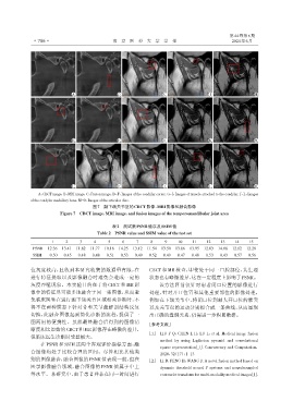


                                A                  D                  G                   J                  M


                                B                  E                  H                   K                  N


                                C                  F                   I                  L                  O
                 A:CBCT image. B:MRI image. C:Fusion image. D-F:Images of the condylar cortex. G-I:Images of muscle attached to the condylar. J-L:Images
              of the condylar medullary bone. M-O:Images of the articular disc.
                                        图7 颞下颌关节区的CBCT影像、MRI影像和融合影像
                           Figure 7 CBCT image,MRI image,and fusion images of the temporomandibular joint area

                                                 表2 测试集PSNR值以及SSIM值
                                          Table 2 PSNR value and SSIM value of the test set

                       1     2     3     4     5     6     7     8     9    10     11    12    13    14    15
               PSNR  12.36  13.41  11.82  11.77  10.18  14.25  13.12  11.54  13.50  13.86  13.95  12.03  14.06  12.02  12.28
               SSIM   0.50  0.45  0.48  0.48  0.51  0.53  0.49  0.52  0.49  0.47  0.48  0.53  0.43  0.57  0.56


              位灰度较高,且收到本研究收集的数据量有限,在                            CBCT 和 MR 检查,即使处于同一口腔部位,其生理
              进行特征提取以及影像融合时难免会造成一定的                             状态也有略微差异,这在一定程度上影响了PSNR。
              灰度吞噬现象。本实验目的在于将 CBCT 和 MR 影                            该方法目前仅针对患者闭口位置的影像进行
              像中的特征尽可能多地融合于同一张图像,从而避                            处理,针对开口位置和其他重要部位的影像处理,
              免临床医生在进行颞下颌关节区域相关诊断时,不                            例如在下颌关节中,将闭口位到最大开口位的髁突
              得不在两种模态下针对骨和关节盘解剖结构反复                             以及关节盘的运动分别拟合成一条曲线,从而显现
              切换,此融合图像起到简化诊断的流程,提供了一                            出正确的盘髁关系,仍需进一步积累数据。
              图两用的便携性。虽然最终融合后得到的图像清
                                                                [参考文献]
              晰度相比原始的CBCT和MR影像存在略微的差异,
                                                                [1] LIU F Q,CHEN L H,LU L,et al. Medical image fusion
              但临床医生诊断时受益极大。
                                                                     method by using Laplacian pyramid and convolutional
                  在 PSNR 和 SSIM 这两个客观评价指标方面,融
                                                                     sparse representation[J]. Concurrency and Computation,
              合图像均处于比较合理的区间。尽管相比其他类
                                                                     2020,32(17):1-13
              别的图像融合,融合图像的PSNR值表现一般,但在
                                                                [2] LI B,PENG H,WANG J. A novel fusion method based on
              医学影像融合领域,融合图像的 PSNR 值属于中上                              dynamic threshold neural P systems and nonsubsampled
              等水平。本研究中,由于患者并非在同一时间进行                                 contourlet transform for multi⁃modality medical images[J].
   45   46   47   48   49   50   51   52   53   54   55