Page 39 - 南京医科大学自然版
P. 39

第45卷第12期        吉澳梅,蒋砚青,韩 振,等. 单侧颞下颌关节不可复性盘前移位与下颌偏斜的相关性研究[J].
                 2025年12月                  南京医科大学学报(自然科学版),2025,45(12):1719-1726,1755                   ·1721 ·


                medicine,DICOM)格式导入 ProPlan CMF 3.0 软件            A             B        B            e
               (Materialise 公司,比利时)。所有CBCT扫描均采用                            c        a
                口腔颌面部专用 CBCT 设备(NewTom/NTVGiEVO,                   Posterior  c’ a’  Anterior          e’  f’  f
                主要技术参数:电压110 kV,电流7.2 mA,扫描时间                                   d          Medial     d
                                                                                                            Lateral
                3.6 s,体素尺寸0.3 mm)。扫描过程中,患者取站立
                位,于牙尖交错位下闭口不动,调整患者头位使眶耳
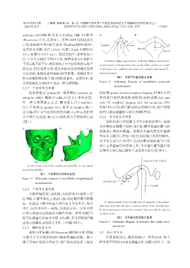
                                                                     A:Median oblique sagittal plane. B:Median oblique coronal plane.
                平面与地平面平行,颅面部的正中矢状面始终与地平                           d:central point of the maximum cross⁃section of the condyle;aa’:anteri⁃
                面垂直,固定患者头部,通过光标定位系统确定患者                           or joint space;cc’:posterior joint space;ee’:superior joint space;ff’:
                头位正确,拍摄患者颅颌面CBCT影像。确保研究中                          lateral joint space.
                所有影像资料均处于相同的照射条件。在软件中,设                                       图2 下颌骨位置测量示意图
                定灰度阈值为500用于重建三维头颅图像。                              Figure 2  Schematic diagram of mandibular positional
                                                                            measurements
                1.2.3 下颌骨形态测量
                    选取鼻根点(nasion,N)、前鼻嵴点(anterior na⁃             加权像(proton density weighted imaging,PDWI)序列
                sal spine,ANS)、蝶鞍点(sella,S)定义正中矢状面。               和快速自旋回波脂肪抑制 T2 加权成像(fast spin
                在三维头颅图像上定点:髁突最上点(condylion,                       echo T2 weighted imaging with fat saturation,FSE
                Cs);下颌角点(gonion,Go);颏下点(menton,Me)。               T2WI⁃FS)序列;闭口斜冠状位PDWI序列;张口带辅
                定义Me至正中矢状面的距离为MS,Cs至Go的距离                         助开口固定装置斜矢状位PDWI序列。
                为下颌升支高度,Me 至 Go 的距离为下颌体部长度                        1.2.6 关节盘状态测量
               (图1)。                                                  每种扫描序列每侧关节有 9 张扫描图片,选择
                                                                  从外侧到内侧最中间的(第5张)髁突截面积最大的
                A                       B                 Cs
                                                                  图像进行观察和测量。患侧关节盘长度及位置测
                                                                  量参考文献[9],在闭口位T2加权像序列进行测量,
                              N
                                                                  关节盘长度为中间带中点到盘最前缘距离与中间
                          S                                       带中点到盘最后缘距离之和,关节盘位置为盘后带
                              ANS                                 后缘中点到长轴与髁突上表面交点的长度(图3)。
                                                            Go

                                         Me                                                    Long axis of the condyle
                        Me
                   A:The frontal view of the maxilla and mandible. B:The lateral
                                                                                            D
                view of the mandible.
                           图1 下颌骨形态测量示意图                                               C
                Figure 1  Schematic diagram of mandibular morphological
                                                                     Articular disc  B
                        measurements
                                                                                A
                                                                                                    Condyle
                1.2.4 下颌骨位置测量
                    分别在轴位面、冠状面、矢状面和 3D 视图上定
                位TMJ,在髁突轴位上逐层扫描,确定髁突最大横截
                面。此截面上髁突的最大直径定义为内外径,取其                               A:anterior border of the articular disc;B:midpoint of the interme⁃
                                                                  diate zone of the articular disc;C:posterior border of the articular disc;
                中点,向外内径作一垂线,形成前后径。以中点所
                                                                  D:intersection point between the condylar long axis and the superior
                在的矢状面及冠状面作为测量界面。参考文献[7]                           condylar surface.
                的方法测量矢状面关节前、后间隙,参考舒凯翔 描                                       图3 关节盘状态测量示意图
                                                          [8]
                述的方法测量冠状面关节外、上间隙(图2)。                             Figure 3  Schematic diagram of articular disc status mea⁃
                                                                           surements
                1.2.5 MRI检查方法
                    使用3.0T核磁(GE Discovery MR750 3.0T)和颞           1.3 统计学方法
                下颌关节专用表面线圈扫描获得 MRI 影像。颞下                              所有指标定点、测量均由同一医师完成,每个
                颌关节MRI扫描序列如下:闭口斜矢状位质子密度                           样本在不同时间点重复测量2次,间隔1周以上。为
   34   35   36   37   38   39   40   41   42   43   44