
Page 60 - 南京医科大学自然版
P. 60
第45卷第12期
·1742 · 南 京 医 科 大 学 学 报 2025年12月
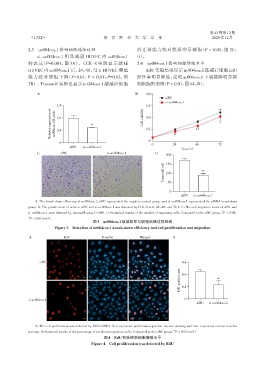
2.3 uc004coz.1影响细胞增殖迁移 的迁移能力较对照组明显减弱(P < 0.01,图 3C、
si⁃uc004coz.1 明显减弱 HUVEC 内 uc004coz.1 D)。
的表达(P=0.001,图 3A)。CCK⁃8 实验显示敲减 2.4 uc004coz.1影响细胞增殖水平
HUVEC 内 uc004coz.1 后,24、48、72 h HUVEC 增殖 EdU 实验结果显示 uc004coz.1 敲减后细胞 EdU
能力较对照组下降(P=0.01,P < 0.01,P=0.02,图 阳性率明显降低,说明 uc004coz.1 下调能够明显抑
3B)。Transwell 实验也显示 uc004coz.1 敲减后细胞 制细胞的增殖(P < 0.01,图4A、B)。
A B 2.0
siNC
si⁃uc004coz.1
1.5 1.5
Relative expression of uc004coz.1/β⁃actin 1.0 ** Cell viability 1.0 ** *
0.5
0.5
0 *
0
0 24 48 72
siNC si⁃uc004coz.1
Time(h)
C siNC si⁃uc004coz.1 D
200
Transwell cell 150 **
100
50
0
siNC si⁃uc004coz.1
A:The knock down efficiency of uc004coz.1;siNC represented the negative control group,and si⁃uc004coz.1 represented the siRNA knockdown
group. B:The growth trend of cells in siNC and si⁃uc004coz.1 was detected by CCK⁃8 at 0,24,48,and 72 h. C:The cell migration levels of siNC and
si⁃uc004coz.1 were detected by transwell assay(×100). D:Statistical results of the number of migrating cells. Compared to the siNC group,P < 0.05,
*
**
P < 0.01(n=3).
图3 uc004coz.1敲减效率与细胞增殖迁移检测
Figure 3 Detection of uc004coz.1 knock⁃down efficiency and cell proliferation and migration
A EdU Hoechst Merged B
siNC 0.6
EdU positive rate 0.4 **
0.2
si⁃uc004coz.1 0
siNC si⁃uc004coz.1
A:The cell proliferation was detected by EdU(×200). Red represents proliferation⁃positive nuclear staining and blue represents nuclear hoechst
**
staining. B:Statistical results of the percentage of proliferative positive cells. Compared to the siNC group,P < 0.01(n=3).
图4 EdU实验检测细胞增殖水平
Figure 4 Cell proliferation was detected by EdU