


Page 69 - 南京医科大学自然版
P. 69
第45卷第6期         瞿  颖,黄    越,李明卉,等. 多模态影像学对乳腺导管原位癌病变范围评估性能的比较[J].
                  2025年6月                     南京医科大学学报(自然科学版),2025,45(6):798-809                        ·807 ·


                      80                                          工智能和放射组学等较为先进、敏感的技术。
                                                           MG     评估的准确性,以在确保手术安全性的基础上,进
                                        *
                 Difference in tumor size between  imaging and histology  -40 0  一步提升保乳手术的成功率,并为临床决策提供重
                             *
                   (mm)
                                                                      本研究旨在提高术前影像学检查对病灶范围
                                                           US
                                         *
                                                   *
                               *
                      40
                                                           MRI
                                                    *
                                                                  要依据。通过系统比较3种影像学检查在病灶范围
                                                                  评估中的效能,本研究得出结论:术前 MRI 成像在
                     -80
                                                                  具有重要价值,建议保乳患者优先接受术前MRI检
                    -120                                          拟行保乳手术的 DCIS/DCIS⁃MI 患者的临床管理中
                             ≤15        16~40       ≥41           查。然而,在基层医疗机构,若缺乏MRI设备,可采
                                    Tumor size(mm)
                                                                  用MG联合US检查作为替代方案,以在一定程度上
                                    *
                                    P < 0.05.
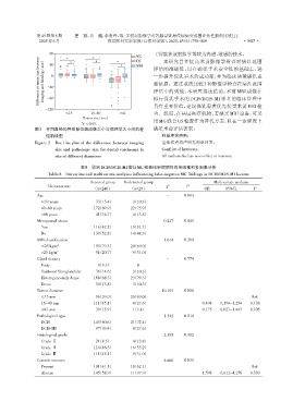
                图2  不同直径的导管原位癌成像大小与病理学大小间的差                       满足术前评估需求。
                    值箱线图                                              利益冲突声明:
                Figure 2  Box line plot of the difference between imaging  全体作者均声明无利益冲突。
                        size and pathologic size for ductal carcinoma in  Conflict of Interests:
                        situ of different diameters                   All authors declare no conflict of interests.
                                   表8 影响DCIS/DCIS⁃MI病灶MG检查结果假阴性的单因素和多因素分析
                     Table 8  Univariate and multivariate analyses influencing false⁃negative MG findings in DCIS/DCIS⁃MI lesions
                                       Detected group  Undetected group                     Multivariate analysis
                     Characteristic                                    χ 2      P
                                        (n=246)         (n=29)                          OR      95%CI        P
                 Age                                                    -      0.863
                   ≤39 years             033(13.4)      03(10.3)
                   40-60 years           172(69.9)      22(75.9)
                   >60 years             041(16.7)      04(13.8)
                 Menopausal status                                     0.217   0.641
                   Yes                   116(47.2)      15(51.7)
                   No                    130(52.8)      14(48.3)
                 BMI classification                                    1.614   0.204
                   ≤25 kg/m 2            195(79.3)      20(69.0)
                   >25 kg/m 2            051(20.7)      09(31.0)
                 Gland density                                          -      0.774
                   Fatty                  08(3.3)          0
                   Scattered fibroglandular  036(14.6)  03(10.3)
                   Heterogeneously dense  168(68.3)     23(79.3)
                   Dense                 034(13.8)      03(10.3)
                 Tumor diameter                                       10.101   0.006
                   ≤15 mm                096(39.0)      20(69.0)                                            Ref.
                   16-40 mm              111(45.1)      08(27.6)                       0.494  0.194-1.254  0.138
                   ≥41 mm                039(15.9)       1(3.4)                        0.175  0.021-1.443  0.105
                 Pathological type                                     1.542   0.214
                   DCIS                  149(60.6)      21(72.4)
                   DCIS⁃MI               097(39.4)      08(27.6)
                 Histological grade                                    2.395   0.302
                   Grade Ⅰ                21(8.5)       04(13.8)
                   Grade Ⅱ               114(46.3)      16(55.2)
                   Grade Ⅲ               111(45.1)      09(31.0)
                 Comedo necrosis                                       4.666   0.031
                   Present               101(41.1)      18(62.1)                                            Ref.
                   Absent                145(58.9)      11(37.9)                       1.598  0.611-4.176  0.339

