


Page 65 - 南京医科大学自然版
P. 65
第45卷第6期         瞿  颖,黄    越,李明卉,等. 多模态影像学对乳腺导管原位癌病变范围评估性能的比较[J].
                  2025年6月                     南京医科大学学报(自然科学版),2025,45(6):798-809                        ·803 ·


                   (cm)  15  r=0.561       (cm)  10  95% LOA -3.54~3.03  (cm)  10 8  r=0.614  (cm)  10  95% LOA -3.15~2.86
                A                     B                         C                     D
                   MG⁃measured tumor size  10 5  Difference in tumor size between MG and histology  -0.25  US⁃measured tumor size  6 4 2  Difference in tumor size between US and histology  -0.14
                                                                      P < 0.001
                      P < 0.001
                                            3.03
                                                                                             2.86
                                           -3.54
                                                                                            -3.15
                    0
                               10
                          5
                     0
                                                                          5
                                                                               10
                       Pathology⁃measured  15  -15  0  5  10  15    0  0  Pathology⁃measured  15  -15  0  5  10  15
                        tumor size(cm)           Mean tumor size(cm)    tumor size(cm)           Mean tumor size(cm)
                E                     F                        G                      H
                   (cm)  15  r=0.853      (cm)  10  95% LOA -1.73~2.44  (cm)  15  r=0.632  (cm)  10  95% LOA -3.17~2.75
                   MRI⁃measured tumor size  10 5  Difference in tumor size between MRI and histology  -1.73  Tumor size measured by MG  combied with US  10 5  Difference in tumor size between MG combined with US and histology  -0.21
                                                                       P < 0.001
                      P < 0.001
                                            2.44
                                                                                             2.75
                                            0.35
                                                                                            -3.17
                    0
                                                                                10
                     0
                          5
                               10
                                                                           5
                       Pathology⁃measured  15  -15  0  5  10  15     0  0  Pathology⁃measured  15  -15  0  5  10  15
                        tumor size(cm)          Mean tumor size(cm)      tumor size(cm)          Mean tumor size(cm)
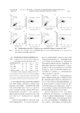
                        图1 3种影像学检查以及MG联合US评估的DCIS/DCIS⁃MI病变范围与病理学大小的相关性和一致性
                Figure 1  Correlation and concordance between DCIS/DCIS ⁃ MI lesion extent and histopathologic size as assessed by the
                        three imaging tests and MG combined US

                  表4 3种影像学检查术前评估病灶范围准确性的比较                        MG联合US检查的准确性与MRI 相当,提示在当前
                Table 4  Comparison of the accuracy of three imaging tests  基层医院中MRI尚未普及并不一定增加保乳术后的
                       for preoperative assessment of lesion extent  二次手术风险,但是由于联合检查的一致性较低,故
                 Examination A vs. B            χ 2      P
                                                                  保乳外观和患者满意度可能受到一定程度影响。多
                                   n01
                                         n10
                 MG vs. US         23    41    5.063    0.033
                                                                  项研究报道了MRI术前评估的潜在价值                 [18-20] ,但也有
                 MG vs. MRI        32    68   12.960   <0.001
                                                                  研究指出    [21] ,MG 在评估 DCIS 病变大小方面与 MRI
                 US vs. MRI        37    57    4.255    0.049
                                                                  无明显差异。然而,这些研究均是回顾性的,用于
                (MG+US)vs. MRI     50    52    0.039    0.921
                                                                  评定影像学大小与病理学大小一致性的标准也各
                   The difference between imaging and pathological lesion size ≤
                5 mm was assigned a value of 0(accurate),and >5 mm was assigned a  不相同,故可能产生上述差异。
                                                                      对成像大小与病理大小一致性的影响因素分
                value of 1(inaccurate). n01 refers to the number of patients where meth⁃
                od A was accurate and method B was inaccurate;n10 refers to the number
                                                                  析提示,肿瘤直径是影响成像大小准确性的重要因
                of patients where method A was inaccurate and method B was accurate.
                                                                  素。故将患者分组进行亚组分析,结果表明对于直
                广泛导管内癌致超声成像边缘不明确;肿瘤体积                             径≥41 mm的导管原位癌,MRI成像范围更准确。因
                较大,图像呈现超出了换能器水平;或受探头宽度                            此,如果计划对病灶广泛的患者行保乳手术,则可
                范围和压力调节的限制。与前述两种影像学检查                             以通过 MRI 高精度评估局部疾病范围。即便对于
                不同,本研究中 MRI“高估”了病灶大小,其原因可                         多中心性的患者,也可通过术前活检和重点评估可
                能是中重度背景实质增强,如肿瘤内导管内成分                             疑病变来规避由此引起的不必要全乳切除。
                或纤维组织成分占比更高             [10,16] 。此外,由于标本              本研究不可避免存在少许不足。首先,本研究
                塌陷和甲醛固定后收缩,组织病理学可能本身就                             为回顾性研究,样本量相对较小,并且数据来自单
                低估了病灶大小        [17] ,故 MRI 或许代表了实际的病              一机构,不可避免样本数据偏倚问题,今后仍需进
                灶范围。                                              一步开展大规模、前瞻性、多中心研究提高结果可信
                    本研究中,MRI 的准确性和一致性也强于其他                        度。其次,本研究由影像科医生读取影像特征并撰写
                两种传统影像学检查。在 DCIS/DCIS⁃MI 患者中,                     报告,可能存在一定的主观偏差,未来应联合应用人

