


Page 42 - 南京医科大学自然版
P. 42
第45卷第7期
               ·940 ·                            南 京    医 科 大 学 学         报                        2025年7月


              A                                      B
                               P < 0.001
                    8
                 The expression of CNN2  (TPM+1)  log2  6 4


                    2


                          Normal     Tumor

                                                                       D
                                                                            1.00                           Low
              C
                                                                                                           High
                   Tumor tissue    Peritumoral tissue
                                                                            0.75
                                                            P < 0.001
                                                      8 6                  Survival probability  0.50


                         200 μm            200 μm   Relative CNN2 expression  4  0.25

                                                      2

                                                                               0 Log⁃rank P=0.004  500  750  1 000
                                                                                      250
                                                      0
                                                         Tumor  Peritumoral                 Time(d)
                                                                           Low 132    70     27     7       2
                                                                           High 88    45     14     3       0
                          50 μm             50 μm
                                                                               0      250    500    750   1 000
                                                                                            Time(d)
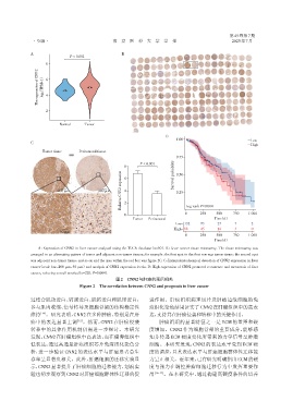
                 A:Expression of CNN2 in liver cancer analyzed using the TCGA database(n=50). B:Liver cancer tissue microarray. The tissue microarray was
              arranged in an alternating pattern of tumor and adjacent non⁃tumor tissues;for example,the first spot in the first row was tumor tissue,the second spot
              was adjacent non⁃tumor tissue,and so on and the area within the red box was figure 2C. C:Immunohistochemical detection of CNN2 expression in liver
              cancer(scale bar=200 μm,50 μm)and analysis of CNN2 expression levels. D:High expression of CNN2 promoted recurrence and metastasis of liver
              cancer, reducing overall survival(n=220,P=0.004).
                                                  图2 CNN2与肝癌的预后相关
                                   Figure 2 The correlation between CNN2 and prognosis in liver cancer


              过结合肌动蛋白、钙调蛋白、肌钙蛋白和肌球蛋白,                           进作用。肝癌组织病理切片及肿瘤边缘细胞的免
              参与肌肉收缩、信号转导及细胞骨架的结构稳定性                            疫组化染色结果证实了CNN2在肝癌组织中的高表
              维持  [14] 。研究表明,CNN2在多种肿瘤,特别是在肝                    达,支持其在肝癌侵袭和转移中的关键作用。
              癌中的表达显著上调          [15] 。然而,CNN2 在肝癌侵袭                 肿瘤组织的显著特征之一是 ECM 的重塑和硬
              转移中的具体作用机制仍需进一步探讨。本研究                             度增加。CNN2 作为细胞骨架的重要成分,能够感
              发现,CNN2在肝癌组织中高表达,而在癌旁组织中                          知并传递 ECM 硬度变化所带来的力学信号至肿瘤
              低表达;通过高通量肝癌组织芯片免疫组化染色分                            细胞。本研究发现,CNN2 的表达水平受到 ECM 硬
              析,进一步验证 CNN2 的表达水平与肝癌患者总生                         度的调控,且其表达水平与肝癌细胞群体性迁移能
              存率呈显著负相关。此外,肝癌细胞的迁移实验显                            力呈正相关。近年来,已有研究明确指出ECM的硬
              示,CNN2显著提升了肝癌细胞的迁移能力,划痕实                          度与张力在调控肿瘤细胞迁移行为中发挥重要作
              验也初步观察到CNN2对肝癌细胞群体性迁移的促                           用 [16-17] 。在本研究中,通过构建高硬度条件的培养

