
Page 88 - 《南京医科大学学报》自然科学版2026年第2期
P. 88
第46卷第2期
·244 · 南 京 医 科 大 学 学 报 2026年2月
表2 3组图像主观评分结果
Table 2 Subjective scoring results of the three sets of images
Subjective evaluation Nread [M(P25,P75)]
Observer F P Pa Pb Pc
indicator Nread⁃128 Nread⁃144 Nread⁃160
Cortical bone depiction Observer1 3(2,3) 3(3,4) 4(3,4) 25.06 <0.001 0.008 <0.001 0.752
Observer2 2(2,3) 3(3,3) 3(3,4) 24.13 <0.001 0.011 <0.001 0.867
Kappa 0.60(0.29,0.92)0.79(0.52,1.00)0.72(0.45,1.00)
Anatomical structure sclarity Observer1 3(2,3) 3(3,4) 4(3,4) 25.09 <0.001 0.006 <0.001 0.993
Observer2 3(2,3) 3(3,4) 4(3,4) 19.85 <0.001 0.102 <0.002 0.555
Kappa 0.62(0.32,0.93)0.65(0.36,0.94)0.76(0.49,1.00)
Perceived image noise Observer1 2(2,3) 3(3,4) 4(3,4) 29.71 <0.001 0.001 <0.001 0.648
Observer2 2(2,3) 3(3,3) 4(3,4) 27.44 <0.001 0.011 <0.001 0.190
Kappa 0.72(0.41,1.00)0.79(0.52,1.00)0.77(0.45,1.00)
Overall image quality Observer1 3(2,3) 3(3,4) 4(3,4) 26.94 <0.001 0.004 <0.001 0.555
Observer2 3(2,3) 3(3,4) 4(3,4) 22.37 <0.001 0.031 <0.001 0.648
Kappa 0.85(0.67,1.00)0.64(0.29,0.99)0.69(0.37,1.00)
Pa:Nread⁃128 vs. Nread⁃144. Pb:Nread⁃128 vs. Nread⁃160. Pc:Nread⁃144 vs. Nread⁃160.
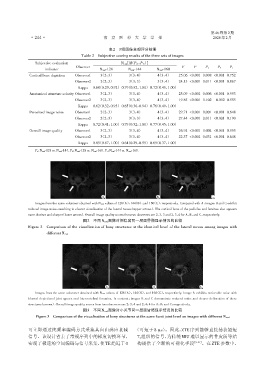
A B C
Images from the same volunteer obtained with N read values of 128(A),144(B),and 160(C),respectively. Compared with A,images B and C exhibit
reduced image noise,resulting in clearer visualization of the lateral recess(upper arrows). The cortical bone of the pedicles and laminae also appears
more distinct and sharper(lower arrows). Overall image quality scores from two observers are 2,3,3 and 2,3,4 for A,B,and C,respectively.
图2 不同Nread图像对侧隐窝同一层面骨骼显示情况的比较
Figure 2 Comparison of the visualization of bony structures at the identical level of the lateral recess among images with
different Nread
A B C
Images from the same volunteer obtained with N read values of 128(A),144(B),and 160(C),respectively. Image A exhibits noticeable noise with
blurred depictionof joint spaces and intervertebral foramina. In contrast,images B and C demonstrate reduced noise and clearer delineation of these
structures(arrows). Overall image quality scores from two observers are 2,3,4 and 2,4,4 for A,B,and C,respectively.
图3 不同Nread图像对小关节同一层面骨骼显示情况的比较
Figure 3 Comparison of the visualization of bony structures at the same facet joint level on images with different N read
可立即通过纯频率编码方式采集其自由感应衰减 (可短于 8 μs)。因此,ZTE 序列能够直接捕获超短
信号。该设计省去了常规序列中的梯度切换环节, T2组织的信号,为传统 MRI 难以显示的骨皮质等结
实现了极速的空间编码与信号采集,使TE趋近于0 构提供了全新的可视化手段 [5,21] 。在 ZTE 参数中,