
Page 26 - 南京医科大学学报自然科学版
P. 26
第43卷第9期
·1204 · 南 京 医 科 大 学 学 报 2023年9月
A 0.6 C D
NC组 HG组 PCDH组 PCDH⁃Rffl组 B # 1.5 1.0 *
0.8
E⁃cadherin 97 kDa 0.4 * 1.0 * # 0.6 #
Fibronectin 263 kDa E⁃cadherin蛋白相对表达量 Fibronectin蛋白相对表达量 α⁃SMA蛋白相对表达量 0.4
α⁃SMA 42 kDa 0.2 0.5 0.2
α⁃Tubulin 50 kDa 0 0 0
PCDH⁃Rffl组
NC组 HG组 PCDH组 NC组 HG组 PCDH组 NC组 HG组 PCDH组
PCDH⁃Rffl组
PCDH⁃Rffl组
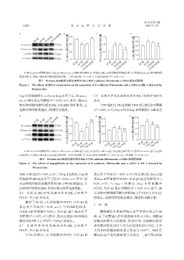
A:Western blot检测各组E⁃cadherin、Fibronectin、α⁃SMA蛋白表达;B:各组E⁃cadherin蛋白表达的定量分析;C:各组Fibronectin蛋白表达的
定量分析;D:各组α⁃SMA蛋白表达的定量分析。与NC组比较,P < 0.05;与PCDH组比较,P < 0.05,n=3。
#
*
图3 Western blot检测过表达Rffl对HK⁃2中E⁃cadherin、Fibronectin、α⁃SMA表达的影响
Figure 3 The effects of Rffl overexpression on the expression of E⁃cadherin,Fibronectin and α⁃SMA in HK⁃2 detected by
Western blot
Dap⁃H组细胞中E⁃cadherin表达水平升高,Fibronec⁃ 2.5 达格列净促进高糖培养的HK⁃2细胞中Rffl 的
tin、α⁃SMA表达均降低(P < 0.05,n=3,图4),提示达 表达
格列净抑制高糖处理的 HK⁃2 的 EMT 和纤维化,且 与NC组相比,HG组细胞中Rffl蛋白表达水平降低
达格列净的浓度越高,抑制作用越强。 (P < 0.05,n=3);Dap⁃L组及Dap⁃H组细胞中Rffl表达
A B 1.5 C D 2.0
NC组 HG组 Dap⁃L组 Dap⁃H组 #△ 1.5 1.5 * #
E⁃cadherin 97 kDa 1.0 # 1.0 * # #△
Fibronectin 263 kDa E⁃cadherin蛋白相对表达量 * Fibronectin蛋白相对表达量 #△ α⁃SMA蛋白相对表达量 1.0
α⁃SMA 42 kDa 0.5 0.5 0.5
α⁃Tubulin 50 kDa 0 0 0
Dap⁃H组
NC组 HG组 Dap⁃L组 NC组 HG组 Dap⁃L组 NC组 HG组 Dap⁃L组
Dap⁃H组
Dap⁃H组
A:Western blot检测各组E⁃cadherin、Fibronectin、α⁃SMA蛋白表达;B:各组E⁃cadherin蛋白表达的定量分析;C:各组Fibronectin蛋白表达的
定量分析;D:各组α⁃SMA蛋白表达的定量分析。与NC组比较,P < 0.05;与HG组比较,P < 0.05;与Dap⁃L组比较,P < 0.05,n=3。
#
*
△
图4 Western blot检测达格列净对HK⁃2中E⁃cadherin、Fibronectin、α⁃SMA表达的影响
Figure 4 The effects of dapagliflozin on the expression of E⁃cadherin,Fibronectin and α ⁃SMA in HK⁃2 detected by
Western blot
均高于HG组(P < 0.05,n=3);与Dap⁃L组相比,Dap⁃H 表达水平升高(P < 0.05,n=3);相比HG组,Dap⁃L组
组细胞中Rffl表达水平升高(P < 0.05,n=3,图5),提 及Dap⁃H组细胞中STAT1、TGF⁃β1表达均降低(P <
示达格列净促进高糖培养的HK⁃2中Rffl的表达,且 0.05,n=3);与 Dap ⁃ L 组 相 比 ,Dap ⁃ H 组 细 胞 中
达格列净的浓度越高,促Rffl表达的作用越明显。 STAT1、TGF⁃β1表达均降低(P < 0.05,n=3,图7),提
2.6 过表达 Rffl 抑制高糖培养的 HK⁃2 细 胞 中 示达格列净抑制高糖培养的HK⁃2中STAT1、TGF⁃β1
STAT1、TGF⁃β1的表达 的表达,达格列净的浓度越高,抑制作用越显著。
相对于 NC 组,HG 组细胞中 STAT1、TGF⁃β1 的
3 讨 论
表达水平升高(P < 0.05,n=3);与 PCDH 组相比,
PCDH⁃Rffl 组细胞中 STAT1、TGF⁃β1 蛋白表达水平 糖尿病是世界范围内日益严重的公共卫生疾
均降低(P < 0.05,n=3,图6),提示过表达Rffl抑制高 病,在全球普通人群中的患病率约为10%。根据国
糖培养的HK⁃2中STAT1、TGF⁃β1的表达。 际糖尿病联合会的数据,到 2035 年,全球糖尿病患
2.7 达 格 列 净 抑 制 高 糖 培 养 的 HK ⁃ 2 细 胞 中 者的数量将从2013年的3.82亿增加到5.92亿,其中
STAT1、TGF⁃β1的表达 大约 40%的糖尿病患者会发展为 DKD 。DKD 是
[8]
与 NC 组相比,HG 组细胞中 STAT1、TGF⁃β1 的 糖尿病最严重的微血管并发症之一,是导致 ESRD