Page 78 - 南京医科大学自然版
P. 78

第44卷第6期
               ·814 ·                            南 京    医 科 大 学 学         报                        2024年6月


              疗科技有限公司的 MR 脑分割软件对所有患者
              3DT1WI图像进行自动勾画(图2),并对每个患者勾
              画图像进行检查并手动修改后,由AI软件自动计算
              出PCSFV和ICV值。
              1.3  统计学方法
                  采用 SPSS 26.0 和 GraphPad Prism 8.3.0 软件进
              行统计分析和作图。统计 ONSD、PCSFV/ICV 和                          The dura can be depicted as the outer hypointense border of the
              CSFP 数据的方差齐性,若数据符合正态分布,采用                         optic nerve sheath(arrows). ONSD is defined as the distance between
                                                                the right and left dural borders(including CSF and optic nerve)at 3 mm
              Pearson 相关分析法分析 CSFP 与 MR 测量结果的相
                                                                behind the papilla(white line).
              关性;反之则采用Spearman相关分析法。然后通过绘                       图1   视神经轴位T2加权MRI显示低信号的视神经被高信
              制受试者工作特征(receiver operating characteristic,            号的脑脊液包围

              ROC)曲线评估 ONSD 和 PCSFV/ICV 联合诊断 CSFP               Figure 1  Axial T2 ⁃ weighted MRI of the orbit indicates
              升高的准确性,并计算曲线下面积(area under curve,                          the hypointense optic nerve is surrounded by
              AUC)。P < 0.05为差异有统计学意义。                                    the hyperintense CSF


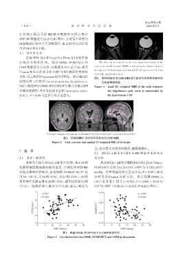
                                 Schematic diagram of peripheral cerebrospinal fluid delineated(pink)and manually modified.
                                            图2 颅脑经轴位、冠状位和矢状位T1加权MRI
                                    Figure 2 Axial,coronal,and sagittal T1⁃weighted MRI of the brain
                                                                主,部分患者出现颈项强直、脑膜刺激征。
              2 结    果
                                                                2.2  NSCLC⁃LM 患者 CSFP 与 MR 测量结果的相关
              2.1  患者一般资料                                       性分析
                  本研究共纳入NSCLC⁃LM患者63例,其中46例                          所有NSCLC⁃LM患者双眼ONSD为(5.23±0.78)mm,

              经脑脊液脱落细胞学检查证实,17 例经典型的 MR                         PCSFV/ICV 比值为 0.21±0.03,CSFP 为(13.82±4.93)
              表现及临床症状确诊,患者年龄(58.08±9.94)岁(范                     mmHg。所有数据均符合正态分布(P > 0.05),相关
              围38~88)岁,男36例(57%),女27例(43%)。63例                  分析采用 Pearson 分析方法。患者双眼 ONSD 与
              患者中伴发脑室增宽20例(32%),脑实质转移32例                        CSFP 呈显著正相关(r=0.567,P < 0.001),PCSFV/
             (51%)。临床症状主要以头晕头痛、恶心、呕吐为                           ICV与CSFP呈负相关(r=-0.365,P=0.003,图3)。

                         7                                         0.30
                             r=0.567                                                         r=-0.365
                            P<0.001                                                           P=0.003
                         6
                       (mm)  5                                     0.25

                       ONSD                                       PCSFV/ICV  0.20

                         4

                         3                                         0.15
                          0    5    10   15   20   25   30             0    5   10   15    20   25   30
                                     CSFP(mmHg)                                   CSFP(mmHg)
                                           图3   患者ONSD、PCSFV/ICV与CSFP的相关性
                                 Figure 3  Correlation between ONSD,PCSFV/ICV and CSFP in all patients
   73   74   75   76   77   78   79   80   81   82   83