


Page 125 - 南京医科大学学报自然科学版
P. 125
第42卷第10期               吴 锦,徐珊珊,张怡帆,等. CT纹理特征评估胰腺导管腺癌的分化程度[J].
                 2022年10月                  南京医科大学学报(自然科学版),2022,42(10):1464-1470,1475                   ·1467 ·


                                                A                         B                        C
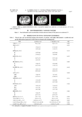
                   A:静脉期CT横断位显示胰腺体尾部低密度肿块,增强后呈相对低密度,伴胰管扩张;B:对病灶最大直径层面的ROI勾画;C:多个ROI经
                重建后获得肿瘤的VOI。
                                          图1 胰腺导管腺癌患者增强CT检查的病灶勾画及重建
                       Figure 1 Focal delineation and reconstruction of ductal adenocarcinoma of the pancreas on enhanced CT

                                     表1 训练组高分化和中⁃低分化PDAC患者临床资料与常规影像特征
                Table 1  Clinical data and conventional imaging characteristics of patients with highly differentiated or moderately and
                        poorly differentiated pancreatic ductal adenocarcinoma in the training group
                             特征                      高分化(n=34)                中⁃低分化(n=32)              P值
                     年龄(岁,x ± s)                      64.2 ± 12.7                62.5 ± 9.6           0.540
                     性别[n(%)]                                                                         0.079
                      男                                15(44.12)                21(65.63)
                      女                                19(55.88)                11(34.37)
                     腹部症状[n(%)]                                                                       0.103
                      是                                19(55.88)                24(75.00)
                      否                                15(44.12)                 8(25.00)
                     胰管扩张[n(%)]                                                                       0.670
                      是                                26(76.47)                23(71.88)
                      否                                8(23.53)                  9(28.12)
                     糖尿病病史[n(%)]                                                                      0.085
                      是                                9(26.47)                 15(46.88)
                      否                                25(73.53)                17(53.12)
                     胆总管扩张[n(%)]                                                                      0.402
                      是                                14(41.18)                10(31.25)
                      否                                20(58.82)                22(68.75)
                     边界[n(%)]                                                                         0.601
                      清楚                               7(20.59)                  5(15.63)
                      模糊                               27(79.41)                27(84.37)
                     血管侵犯[n(%)]                                                                       0.510
                      有                                11(32.35)                 8(25.00)
                      无                                23(67.65)                24(75.00)
                     肿瘤位置[n(%)]                                                                       0.720
                      头颈                               14(41.18)                15(46.87)
                      钩突                               4(11.76)                  5(15.63)
                      体尾                               16(47.06)                12(37.50)
                     胰腺萎缩[n(%)]                                                                       0.880
                      是                                9(26.47)                  9(13.64)
                      否                                25(73.53)                23(71.87)
                     肿瘤大小(cm,x ± s)                   2.56 ± 0.93               2.95 ± 1.20           0.226
                     平扫CT值(HU,x ± s)                  29.82 ± 8.72              29.63 ± 5.67          0.914
                     TPER[HU,M(P25,P75 )]           0.62(0.41,0.77)           0.46(0.39,0.60)         0.005
                     CA19⁃9[U/mL,M(P25,P75 )]      99.28(13.46,442.90)      101.53(24.38,437.70)      0.546
                     CA125[U/mL,M(P25,P75 )]        7.80(0.85,2.87)           13.80(0.73,2.98)        0.001
                   TPER:肿瘤实质与正常胰腺组织的静脉期CT值的比值(tumor to pancreas ratio at portal venous phase)。

