


Page 57 - 南京医科大学自然版
P. 57
第44卷第9期           郭   琼,唐娜娜,焦春花,等. 气囊辅助式小肠镜在诊断Meckel憩室中的应用价值[J].
                  2024年9月                     南京医科大学学报(自然科学版),2024,44(9):1227-1231                      ·1229 ·


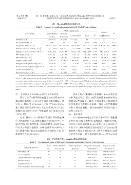
                                                   表1 Meckel憩室合并并发症分析
                                   Table 1 Analysis of complications associated with Meckel’s diverticulum

                                                              With complications
                                                                                                 Without
                         Clinical data      Gastrointestinal  Intestinal                                    P c
                                                                             a             b
                                                                    Diverticulitis  Total  P   complications
                                               bleeding   obstruction
                Numbers[n(%)]                 .30(54.5)   14(25.5)0  11(20)0.0.    55      -       32        -
                Sex(male/female,n)              24/6        9/5         8/3      41/14   0.545    20/12    0.273
                Age[years,M(P25,P75)]        26.5(17.8,41) 38(17,67.3) 38(19,64).  29(18,52) 0.288 49.5(29.3,63) 0.003
                Disease duration[d,M(P25,P75)]  0.07(3,155.8) 04(2,8)0.0  007(1,158.8)  7(2,60) 0.339  -     -
                Distance to ileocecal valve(cm,x ± s)  76.9 ± 25.2  62.5 ± 19.7  62.2 ± 31.5  70.2 ± 25.6 0.138  74.3 ± 28.8  0.479
                MD length(cm,x ± s)            5.6 ± 2.5  5.6 ± 3.0   3.4 ± 1.5  5.2 ± 2.6  0.070  3.3 ± 1.4  0.001
                MD width(cm,x ± s)             2.3 ± 1.0  2.6 ± 1.4   2.4 ± 0.9  2.4 ± 1.1  0.819  1.6 ± 0.7  0.002
                Surgical rate[n(%)]           .25(83.3)   14(100.0)  10(90.9)0  49(89.1) 0.316  0.18(56.3) <0.001
                Preoperative diagnosis rate[n(%)]  .19(76.0)  1(7.1)0  1(10.0)  21(42.9) 0.000 d  0(0)     0.001
                Ectopic tissues on pathology[n(%)]  .11(42.3)  2(14.3)  0(0)0.0  13(26.0) 0.019 e  0.02(13.3)  0.488
                Gastric tissue[n(%)]          .11(42.3)    0(0)0.0    0(0)0.0   11(22.0) 0.001 f  0(0)     0.055
                Pancreatic tissue[n(%)]         0(0)0      2(14.3)    0(0)0.0    2(4.0)  0.111  0.02(13.3)  0.226
                   a:Diverticulitis(combined gastrointestinal perforation or not);b:P⁃value comparing intestinal obstruction,gastrointestinal bleeding,and diverticu⁃
                litis;c:P⁃value comparing the groups with and without complications;d:The gastrointestinal bleeding and intestinal obstruction groups differed from
                one another after multiple comparisons using Bonferroni’s method(P < 0.05);e:The gastrointestinal bleeding and diverticulitis groups differed from
                one another after multiple comparisons using Bonferroni’s method(P < 0.05);f:There was a difference between the groups with gastrointestinal bleed⁃
                ing and intestinal obstruction(P < 0.05)and gastrointestinal bleeding and diverticulitis(P < 0.05)after multiple comparisons using the Bonferroni’s
                method.

                2.3  不同检查方法对Meckel憩室的诊断价值                             由此可见,气囊辅助式小肠镜对Meckel憩室的
                    研究评估了6种不同的检查方法对小肠Meckel                       诊断灵敏度最高,仅有 2 例因进镜困难未能顺利达
                憩室的诊断价值,计算各种方法的诊断灵敏度。结                            到憩室位置而漏诊。此外,15例患者在小肠镜检查
                果如下:腹部 CT 8.2%(4/49);小肠 CT 9.4%(3/32);            中发现憩室旁或憩室内溃疡,1 例见异位胃黏膜样
                数字减影血管造影 0(0/9);核素显像 58.3%(7/12);                 结构,1 例见黏膜桥,1 例见活动性出血并行内镜下
                胶囊内镜 30.0%(3/10);气囊辅助式小肠镜 94.4%                   钛夹止血治疗。
               (34/36,表2)。                                        2.4  临床结局
                    此外,腹部 CT 有 4 例提示不明原因的肠壁增                          有67例Meckel憩室患者接受手术治疗,腹腔镜
                厚、1例肠腔内占位,诊断获益率为18.4%(9/49);小                     手术16例,开腹手术51例,包括急诊开腹探查21例,
                肠CT 发现2例肠壁增厚、1例肠腔狭窄、4例腔内占                         术前仅21例明确诊断Meckel憩室。另外20例未手
                位以及 1 例肠套叠梗阻,诊断获益率为 34.4%(11/                     术患者中,14例为偶然发现的无并发症的Meckel憩
                32);胶囊内镜 2 例发现回肠溃疡、1 例腔内占位,诊                      室患者,5例合并消化道出血,1例合并憩室炎,经保
                断获益率为60.0%(6/10)。                                 守治疗后好转。对手术切除的 Meckel 憩室标本进

                                              表2  不同检查方法对Meckel’s憩室诊断的价值
                             Table 2  The diagnostic value of different examination methods for Meckel’s diverticulum

                                             Total  Certain Meckel’s  Other significant  Diagnostic sensitivity  Diagnostic yield
                        Different methods
                                             (n)     diverticulum    findings     [%(95%CI)]       [%(95%CI)]
                  Abdominal CT                49         04             5          8.2(2.3-19.6)  18.4(8.8-32.0)0
                  CT enterography             32         03             8          9.4(2.0-25.0)  34.4(18.6-53.2)
                  Digital subtraction angiography  09    00             0           .0(0-33.6 )      0(0-33.6)
                  Nuclide imaging             12         07             0         58.3(27.7-84.8)  58.3(27.7-84.8)
                  Capsule endoscopy           10         03             3         30.0(6.7-65.2)0  60.0(26.2-87.8)
                  Balloon⁃assisted small enteroscopy  36  34            0         94.4(81.3-99.3)  94.4(81.3-99.3)

