Page 63 - 南京医科大学自然版
P. 63

第45卷第10期          任言晖,徐可颖,张 伟,等. 成纤维细胞来源半乳糖凝集素⁃3通过细胞外基质蓄积促进矽
                 2025年10月               肺纤维化[J]. 南京医科大学学报(自然科学版),2025,45(10):1427-1434                  ·1431 ·


                沉积,表现为鲜红色细网状分布模式(图2A)。偏振                          A
                                                                              NS                    SiO2
                光显微镜观察进一步证实,SiO2暴露组肺泡壁区域
                Ⅰ型胶原显著增多增厚,呈现特征性的亮红色及明
                黄色双折射现象,而NS组仅在血管壁周围观察到少
                量Ⅰ型胶原分布(图 2B)。组织染色结合偏振光分
                析结果表明,SiO2暴露导致小鼠肺组织胶原成分发                                                                    50 μm
                生显著改变,表现为胶原结构重构及比例失调,呈                            B           NS
                现典型的肺纤维化病理特征。上述实验结果证实                                                               SiO2
                SiO2诱导的小鼠肺纤维化模型成功建立。

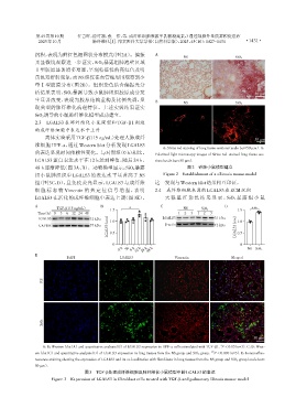
                2.3  LGALS3 在肺纤维化小鼠模型和 TGF⁃β1 刺激
                的成纤维细胞中表达水平上升
                    离体实验采用TGF⁃β1(5 ng/mL)处理人肺成纤
                                                                                                            50 μm
                维细胞 HPF⁃a,通过 Western blot 分析发现 LGALS3
                                                                     A:Sirius red staining of lung tissue sections(scale bar=50 μm). B:
                的表达呈现时间依赖性变化。与对照组(0 h)相比,                         Polarized light microscopy images of Sirius red⁃stained lung tissue sec⁃
                LGALS3 蛋白表达水平在 12 h 达到峰值,随后 24 h、                 tions(scale bar=50 μm).
                48 h逐渐降低(图3A、B)。动物模型显示:SiO2暴露                                   图2 矽肺小鼠模型建立
                                                                     Figure 2  Establishment of a silicosis mouse model
                组小鼠肺组织中 LGALS3 的表达水平显著高于 NS
                组(图 3C、D),且免疫荧光显示,LGALS3 与成纤维                     这一发现与Western blot结果相互印证。
                细 胞 标 志 物 Vimentin 的 共 定 位 信 号 增 强 ,表 明           2.4  成纤维细胞来源的LGALS3在ECM沉积
                LGALS3 在活化的成纤维细胞中表达上调(图 3E)。                          天 狼 星 红 染 色 结 果 显 示 ,SiO2 暴 露 组 小 鼠

                A                          B                        C                          D
                         TGF⁃β1(5 ng/mL)               *                      NS                           ***
                                               1.5                                    SiO2           1.5
                 Time(h) 0  3  6 12 24 48                                   1  2  3  1  2  3
                                              level  1.0                                            level  1.0
                 LGALS3                31 kDa                        LGALS3                31 kDa
                                              LGALS3  0.5                                           LGALS3  0.5
                 GAPDH                 37 kDa                         β⁃actin              42 kDa

                                                0                                                     0
                                                   0 h  3h  6 h  12 h  24 h  48 h                        NS SiO2
                E
                             DAPI                  LGALS3                 Vimentin                Merged


                   NS


                   SiO2


                                                                                                            50 μm
                                                                                              *
                   A,B:Western blot(A)and quantitative analysis(B)of LGALS3 expression in HPF⁃a cells stimulated with TGF⁃β1. P < 0.05(n=3). C,D:West⁃
                ern blot(C)and quantitative analysis(D)of LGALS3 expression in lung tissues from the NS group and SiO 2 group.  *** P < 0.001(n=3). E:Immunofluo⁃
                rescence staining showing the expression of LGALS3 and its co⁃localization with fibroblasts in lung tissues from the NS group and SiO 2 group(scale bar=
                50 μm).
                                     图3 TGF⁃β处理成纤维细胞及肺纤维化小鼠模型中的LGALS3的表达
                       Figure 3  Expression of LGALS3 in fibroblast cells treated with TGF⁃β and pulmonary fibrosis mouse model
   58   59   60   61   62   63   64   65   66   67   68