Page 64 - 南京医科大学自然版
P. 64

第45卷第10期
               ·1432 ·                           南 京    医 科 大 学 学         报                        2025年10月


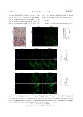
              ECM 中胶原沉积增多且结构致密(图 4A)。组织                         水平上升(图 4D、E);移除种植的细胞后,实验组
              免 疫 荧 光 染 色 显 示 ,与 NS 组 相 比 ,SiO2 暴 露 组           ECM上的LGALS3表达水平仍高于对照组(图4F、G)。
              ECM 中 LGALS3 的蓄积显著增加(图 4B、C)。将
                                                                3  讨 论
              TGF⁃β1 刺激的小鼠肺成纤维细胞 MLg 种植于对
              照组小鼠肺组织提取的 ECM 上后,LGALS3 表达                            矽肺作为进行性纤维化性间质性肺疾病(pro⁃
                 A                           B                                               C
                                                         DAPI              LGALS3
                                                                                                (%)  5 4  **

                    NS                           NS                                             positive area  3 2

                                                                                                LGALS3  1 0


                                                                                                    NS SiO2
                    SiO2                         SiO2


                                   200 μm                                          50 μm
                    D                                                                    E
                                DAPI                LGALS3               Merged
                                                                                             (%)  6  *
                                                                                             positive area  4
                       NS

                                                                                             LGALS3 2
                                                                                  50 μm       0
                                                                                                PBS TGF⁃β1

                       SiO2


                                                                                  50 μm

                    F                                                                    G
                                DAPI                LGALS3               Merged
                                                                                             (%)  6  *

                       NS                                                                    positive area  4

                                                                                             LGALS3 2
                                                                                  50 μm
                                                                                              0
                                                                                                PBS TGF⁃β1

                       SiO2


                                                                                  50 μm

                 A:Sirius red staining of the extracellular matrix(ECM)in the NS group and SiO2 group(scale bar=200 μm). B,C:Immunofluorescence staining
                                                                                                    **
             (B)and quantitative analysis(C)showing the expression of LGALS3 on the ECM in the NS group and SiO 2 group(scale bar=50 μm),P < 0.01(n=3).
              D,E:Immunofluorescence staining(D)and quatitative analysis(E)showing the expression of LGALS3 on the ECM seeded with MLg cells treated with
                                      *
              PBS or TGF⁃β1(scale bar=50 μm),P < 0.05(n=3). F,G:Immunofluorescence(F)and quatitative analysis(G)staining showing the expression of
                                                                           *
              LGALS3 on the ECM after removal of MLg cells treated with PBS or TGF⁃β1(scale bar=50 μm). P < 0.05(n=3).
                                                图4 小鼠ECM上LGALS3蓄积情况
                                         Figure 4  Accumulation of LGALS3 on the ECM in mice
   59   60   61   62   63   64   65   66   67   68   69