


Page 114 - 南京医科大学自然版
P. 114
第45卷第11期
               ·1644 ·                           南 京    医 科 大 学 学         报                        2025年11月


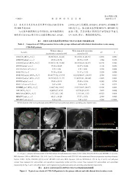
              2.2  未发生与发生临床恶化事件CTD⁃PAH患者组                       LVSVI、LVCI 均减低,RVEDVI、RVESVI、RVMMI 和
              间CMR参数比较                                          VMI 均升高。肺动脉血流参数 MPAPV、MPAMV 均
                  与未发生临床恶化事件组相比,发生临床恶化                          显著下降。其余参数在两组间差异无统计学意义
              事件组 CTD⁃PAH 患者的心功能参数 LVEF、RVEF、                  (P > 0.05,表1)。典型病例见图2。

                                    表1 未发生与发生临床恶化事件组CTD⁃PAH患者CMR参数比较
                  Table 1 Comparison of CMR parameters between the groups without and with clinical deterioration events among
                         CTD⁃PAH patients

                                                Without clinical    With clinical deterioration
                    Variable                                                                  t/Z          P
                                            deterioration events(n=99)   events(n=37)
                LVEF[%,M(P25,P75 )]           58.56(52.41,63.28)      54.18(43.10,60.71)    -2.051       0.040
                LVEDVI(mL/m ,x ± s)             65.96 ± 19.52            60.78 ± 14.95       1.462       0.146
                           2
                LVESVI[mL/m ,M(P25,P75 )]     27.00(21.30,33.86)      28.18(22.43,36.27)    -0.570       0.569
                           2
                LVSVI(mL/m ,x ± s)              37.02 ± 11.85            32.27 ± 11.06       2.116       0.036
                          2
                LVCI[L/(min·m),x ± s]            2.76 ± 0.84             2.31 ± 0.66         2.924       0.004
                            2
                RVEF(%,x ± s)                   37.54 ± 12.84            31.97 ± 13.32       2.228       0.028
                RVEDVI[mL/m ,M(P25,P75 )]     89.40(77.52,115.53)    112.92(86.47,134.93)   -2.589       0.010
                            2
                RVESVI[mL/m ,M(P25,P75 )]     54.79(42.15,71.77)      71.06(53.80,109.04)   -3.005       0.003
                           2
                RVSVI(mL/m ,x ± s)              35.69 ± 13.93            35.00 ± 12.36       0.262       0.794
                          2
                RVCI[L/(min·m),x ± s]            2.67 ± 0.99             2.50 ± 0.72         1.104       0.273
                            2
                RVMMI[g/m ,M(P25,P75 )]       11.06(7.58,15.51)       15.27(10.57,20.17)    -2.920       0.003
                         2
                VMI[M(P25,P75 )]               0.24(0.17,0.39)         0.37(0.25,0.55)      -3.055       0.002
                MPA/AAod[M(P25,P75 )]          1.17(1.03,1.36)         1.19(1.04,1.33)      -0.523       0.601
                MPAPV(cm/s,x ± s)               68.13 ± 19.26            57.57 ± 15.51       2.989       0.003
                MPAMV(cm/s,x ± s)                8.26 ± 3.22             5.81 ± 2.23         4.260      < 0.001
                 MPAd:diameter of the main pulmonary artery;MPA/AAod:main pulmonary artery to ascending aorta diameter ratio.


                                                                                                Velocity vs Time
                                                                                          cm/sec       legend
                                                                                        53               Date
                                                                                                         Spine(+/- 1)
                                                                                        47
                                                                                        41
                                                                                        35
                                                                                        29
                                                                                        23
                                                                                        17
                                                                                        11
                                                                                                           Time
                                                                                         5                (ms)
                                                                                        -1
                                                                                         0  82 164 246 328 410 492 574 656 738 820
                                                                                        -7
                                    A                        B                       C  Slice position:SP H64.7 Venc adjustment -110∶110  D
                                                                                       Check contours,computer generated contours may not correspond to anatomy.
                                                                                                Velocity vs Time
                                                                                          cm/sec       legend
                                                                                        26               Date
                                                                                                         Spine(+/- 1)
                                                                                        23
                                                                                        20
                                                                                        17
                                                                                        14
                                                                                        11
                                                                                         8
                                                                                         5
                                                                                         2                 Time
                                                                                                           (ms)
                                                                                        -1 0  71 142 213 284 355 426 497 568 639 710
                                                                                        -4
                                    E                        F                       G  Slice position:SP P77.7 Venc adjustment -70∶70  H
                                                                                       Check contours,computer generated contours may not correspond to anatomy.
                 A-D:Case 1,a 44⁃year⁃old woman with CTD⁃PAH without clinical deterioration events. RVEF:54%;RVEDVI:85.85 mL/m ;MPAMV:11.02 cm/s;
                                                                                               2
              MPA diameter:3.08 cm;MPA/AAod:1.28. E-H:Case 2,a 34⁃year⁃old woman with CTD⁃PAH who experienced disease progression requiring rehospita⁃
                                              2
              lization. RVEF:16.5%;RVEDVI:225.32 mL/m ;MPAMV:4.36 cm/s;MPA diameter:3.86 cm;MPA/AAod:1.59. In Fig. A and E,red and green
              contour lines represent LV endocardium and epicardium respectively ;yellow and blue contour lines represent RV endocardium and epicardium
              respectively. In Fig. C and G,the yellow lines 1 and 2 represent main pulmonary artery and ascending aorta diameter respectively.
                                     图2 未发生与发生临床恶化事件组CTD⁃PAH患者的典型病例图
                   Figure 2  Typical case charts of CTD⁃PAH patients in the groups without and with clinical deterioration events

