


Page 61 - 南京医科大学自然版
P. 61
第45卷第5期          孙书凝,龚予希,杨野梵,等. CYB5R2在弥漫大B细胞淋巴瘤中的表达及其临床意义[J].
                  2025年5月                     南京医科大学学报(自然科学版),2025,45(5):644-651                        ·647 ·


                             HE                     IHC                     HE                     IHC


                 DLBCL


                                        A                       B                      C                      D


                 FL


                                        E                       F                      G                      H


                 MALT


                                        I                       J                      K                       L


                 MCL


                                        M                       N                      O                       P


                 SLL/CLL


                                        Q                       P                      S                       T
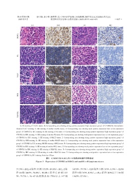
                   A:HE staining of DLBCL tissue. B:Corresponding area showing strong positive expression(high expression group)of CYB5R2 by immunohisto⁃
                chemical(IHC)staining. C:HE staining of another DLBCL tissue. D:Corresponding area showing weak positive expression(zero or low expression
                group)of CYB5R2 by IHC staining. E:HE staining of FL tissue. F:Corresponding area showing strong positive expression(high expression group)of
                CYB5R2 by IHC staining. G:HE staining of another FL tissue. H:Corresponding area showing weak positive expression(zero or low expression group)
                of CYB5R2 by IHC staining. I:HE staining of MALT tissue. J:Corresponding area showing strong positive expression(high expression group)of
                CYB5R2 by IHE staining. K:HE staining of another MALT tissue. L:Corresponding area showing weak positive expression(zero or low expression
                group)of CYB5R2 by IHC staining. M:HE staining of MCL tissue. N:Corresponding area showing strong positive expression(high expression group)of
                CYB5R2 by IHC staining. O:HE staining of another MCL tissue. P:Corresponding area showing weak positive expression(zero or low expression group)
                of CYB5R2 by IHC staining. Q:HE siaining of SLL/CLL tissue. R:Corresponding area showing strong positive expression(high expression group)of
                CYB5R2 by IHC staining. S:HE staining of another SLL/CLL tissue. T:Corresponding area showing weak positive expression(zero or low expression
                group)of CYB5R2 by IHC staining. Scale bar=25μm.
                                           图2 CYB5R2在DLBCL和小B细胞淋巴瘤中的性表达
                                 Figture 2 Expression of CYB5R2 in DLBCL and small B⁃cell lymphoma tissues


                37.3%),BCL⁃6 阳性 53 例(53/59,89.8%),BCL⁃2 阳        (47/59,79.7%),CD5 阳性 5 例(5/59,8.5%),CD30

                性 46 例(46/59,78.0%),MUM⁃1 阳性有 47 例(47/            阳性 5 例(5/59,8.5%),c⁃Myc 阳性(40%以上)16 例
                59,79.7%),Ki⁃67 高增殖表达(75%以上)47 例                 (16/59,27.1%)。

