


Page 10 - 南京医科大学自然版
P. 10
第46卷第8期
               ·1116 ·                           南 京    医 科 大 学 学         报                        2026年8月


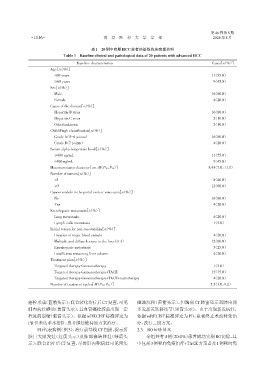
                                           表1 20例中晚期HCC患者的基线临床病理资料
                              Table 1 Baseline clinical and pathological data of 20 patients with advanced HCC

                                    Baseline characteristics                                       Cases[n(%)]
                        Age[n(%)]
                          <60 years                                                              0.11(55.0)
                          ≥60 years                                                              0.09(45.0)
                        Sex[n(%)]
                          Male                                                                   0.16(80.0)
                          Female                                                                 0.04(20.0)
                        Cause of the disease[n(%)]
                          Hepatitis B virus                                                      0.16(80.0)
                          Hepatitis C virus                                                      0.02(10.0)
                          Other/unknown                                                          00.2(10.0)
                        Child⁃Pugh classification[n(%)]
                          Grade A(5-6 points)                                                    0.16(80.0)
                          Grade B(7 points)                                                      0.04(20.0)
                        Serum alpha⁃fetoprotein level[n(%)]
                          ≥400 ng/mL                                                             0.11(55.0)
                          <400 ng/mL                                                             0.09(45.0)
                        Maximum tumor diameter[cm,M(P25,P75)]                                    8.45(7.0,11.1)
                        Number of tumors[n(%)]

                          <3                                                                     0.08(40.0)
                          ≥3                                                                     0.12(60.0)
                        Cancer emboli in the portal vein or vena cava[n(%)]
                          No                                                                     0.16(80.0)
                          Yes                                                                    0.04(20.0)
                        Extrahepatic metastasis[n(%)]
                          Lung metastasis                                                        0.04(20.0)
                          Lymph node metastasis                                                  0.01(5.0)
                        Initial reason for non⁃resectability[n(%)]
                          Invasion of major blood vessels                                        0.04(20.0)
                          Multiple and diffuse lesions in the liver(≥ 3)                         0.12(60.0)
                          Extrahepatic metastasis                                                0.05(25.0)
                          Insufficient remaining liver volume                                    0.04(20.0)
                        Treatment plan[n(%)]
                          Targeted therapy+immunotherapy                                         0.01(5.0)
                          Targeted therapy+immunotherapy+TACE                                    0.15(75.0)
                          Targeted therapy+immunotherapy+TACE+radiotherapy                       0.04(20.0)
                        Number of treatment cycles[M(P25,P75)]                                   05.5(3.0,9.0)


              癌栓形成(蓝箭头示);联合转化治疗后CT复查,可见                         碘油沉积(黄箭头示),但胸部CT肺窗显示双肺出现
              肝内病灶碘油(黄箭头示),且血管癌栓较前出现一定                          多发新发转移结节(黑箭头示)。由于出现新发病灶,
              程度的退缩(紫箭头示)。依据mRECIST标准评定为                        依据mRECIST标准评定为PD,患者终止术前转化治
              PR但未达手术指针,患者继续维持原方案治疗。                            疗,改行二线方案。
                  PD代表病例(图3):治疗前基线CT扫描,提示肝                      2.3 R0切除情况
              脏巨大原发灶(红箭头示)及肺部寡转移灶(绿箭头                                全组共有4例(20.0%)患者成功实现R0切除,其
              示);联合治疗后CT复查,尽管肝内靶病灶可见密集                          中包括3例靶向免疫治疗+TACE方案者及1例靶向免

