
Page 36 - 南京医科大学学报自然科学版
P. 36
第41卷第4期
·506 · 南 京 医 科 大 学 学 报 2021年4月
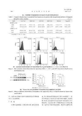
表2 肝癌切除术中洛铂腹腔灌注治疗患者外周血淋巴细胞表型的变化
Table 2 Changes in the phenotype of peripheral blood lymphocytes in patients with intraperitoneal perfusion of lobaplatin
during liver cancer resection (x ± s)
组别 CD3(%) CD4(%) CD8(%) CD4 /CD8 + NK(%) Treg(%)
+
+
+
+
洛铂组(n=40)
术前 66.37 ± 6.73 35.34 ± 7.06 28.11 ± 8.66 1.48 ± 0.86 17.92 ± 7.77 9.76 ± 2.86
术后 63.92 ± 11.48 38.50 ± 7.40 * 24.42 ± 12.04 2.28 ± 1.76 ** 20.59 ± 10.06 8.81 ± 1.58 *
对照组(n=33)
术前 65.94 ± 6.84 34.18 ± 8.11 29.06 ± 6.44 1.14 ± 0.53 17.67 ± 8.03 10.11 ± 2.81
术后 64.94 ± 10.28 33.63 ± 11.54 27.73 ± 8.23 1.39 ± 0.78 20.04 ± 10.69 09.51 ± 1.67
与术前比较,P < 0.05,P < 0.01。
*
**
TAP⁃1 TAP⁃2 HLA⁃Ⅰ
1 500 32.4% 1 500 43.8% 1 500 27.9%
1 000 1 000 1 000
对照组
500 500 500
0 0 0
0 10 2 10 3 10 4 10 5 0 10 2 10 3 10 4 10 5 0 10 2 10 3 10 4 10 5
1 500 94.4% 1 500 97.3% 1 500 54.0%
1 000 1 000 1 000
洛铂组
500 500 500
0 0 0
2 3 4 5 2 3 4 5 2 3 4 5
0 10 10 10 10 0 10 10 10 10 0 10 10 10 10
图1 流式细胞分析显示洛铂组(LBP)相比对照组(PBS)Hep3B肝癌细胞HLA⁃Ⅰ、TAP⁃1、TAP⁃2表达升高
Figure 1 Flow cytometry analysis showed that the expressions of HLA⁃Ⅰ,TAP⁃1,and TAP⁃2 increased in Hep3B cells in
the lobaplatin group
PD⁃L1蛋白相对表达量 PD⁃L1 mRNA相对表达量
洛铂组(μmol/L) 2.5 2.5 **
对照组 8 8 16 16 2.0 ** 2.0 *
PD⁃L1 40~50 kDa 1.5 * 1.5
GAPDH 37 kDa 1.0 1.0
0.5
0.5
0.0
对照组 8 16 0.0 对照组 8 16
洛铂组(μmol/L) 洛铂组(μmol/L)
两组比较,P < 0.05,P < 0.01(n=3)。
*
**
图2 Western blot、RT⁃qPCR检测不同浓度洛铂对Hep3B细胞PD⁃L1表达影响
Figure 2 Effects of different concentrations of lobaplatin on the expression of PD⁃L1 in Hep3B detected by Western blot
and RT⁃qPCR
[3]
4)。说明 AKT 通路可能参与调控洛铂对肝癌细胞 速、术后复发率高导致患者长期生存率较低 。治
PD⁃L1表达的影响。 疗手段包括手术治疗(肝切除术和肝移植)、局部消
融、TACE、放射治疗以及全身治疗(系统化疗、分子
3 讨 论
靶向治疗、免疫治疗等),肝癌传统化疗虽在临床取
肝癌因起病隐匿、早期诊断困难、病情进展迅 得一定疗效,但患者获益有限。既往研究表明肝癌