
Page 105 - 南京医科大学学报自然科学版
P. 105
第44卷第3期 陈 磊,邹月芬,祁 良. 冠状位T2⁃weighted Dixon成像在棘间韧带退变分型中的应用价值[J].
2024年3月 南京医科大学学报(自然科学版),2024,44(3):393-397 ·395 ·
A B C D E F
G H I J K L
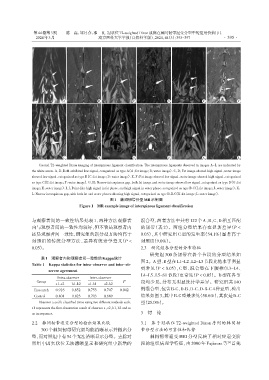
Coronal T2⁃weighted Dixon imaging of interspinous ligament classification. The interspinous ligaments observed in images A-L are indicated by
the white arrow. A,B:Both exhibited low signal,categorized as type A(A:fat image;B:water image). C,D:Fat image showed high signal,water image
showed low signal,categorized as type B(C:fat image;D:water image). E,F:Fat image showed low signal,water image showed high signal,categorized
as type C(E:fat image;F:water image). G,H:Narrow interspinous gap,both fat image and water image showed low signal,categorized as type D(G:fat
image;H:water image). I,J:Point⁃like high signal in fat phase,and high signal in water phase,categorized as type B⁃C(I:fat image;J:water image). K,
L:Narrow interspinous gap,with both fat and water phases showing high signal,categorized as type D⁃B⁃C(K:fat image;L:water image).
图1 棘间韧带分型MR示例图
Figure 1 MR example image of interspinous ligament classification
与观察者间的一致性结果见表 1,两种方法观察者 混合型,两者方法中共有122个A、B、C、D相互匹配
内与观察者间的一致性均较好,但不管是观察者内 的韧带(表 2)。两组分型结果存在显著差异(P <
还是观察者间一致性,研究组的新分型方法均高于 0.05),其中研究组 C 型的发生率(54.1%)显著高于
对照组的传统分型方法,差异有统计学意义(P < 对照组(9.0%)。
0.05)。 2.3 研究组各分型的分布特征
研究组 300 条韧带在各个节段的分型结果如
表1 观察者内和观察者间一致性的Kappa统计
Table 1 Kappa statistics for intra⁃observer and inter⁃ob⁃ 图 2。A型、B型在L1~L2、L2~L3节段比他水平明显
server agreement 更常见(P < 0.05),C 型、混合型在下腰椎(L3~L4、
L4~L5、L5~S1节段)更常见(P < 0.05)。D型在各节
Intra⁃observer Inter⁃observer
Group P 段均少见,分布无明显统计学差异。研究组共 140
a1⁃a2 b1⁃b2 a1⁃b1 a2⁃b2
例混合型,包含B⁃C、D⁃B、D⁃C、D⁃B⁃C 4种亚型,检出
Research 0.916 0.852 0.753 0.747 0.042
Control 0.801 0.823 0.703 0.689 结果如图3,其中B⁃C型最多见(58.6%),其次是D⁃C
Observer a and b classified twice using two different methods each. 型(25.0%)。
a1 represents the first observation result of observer a,a2,b1,b2 and so
on in sequence. 3 讨 论
2.2 棘间韧带退变分型的检出结果比较 3.1 基于冠状位 T2⁃weighted Dixon 序列的棘间韧
300个棘间韧带研究组均能清晰显示并做出分 带分型方法的可靠性和优势
型,而对照组中有54个无法清晰显示分型。去除对 棘间韧带退变 MRI 分型反映了相对应退变阶
[6]
照组中因矢状位无法清晰显示和研究组中新增的 段的组织病理学特征,由 2000 年 Fujiwara 等 首先