
Page 36 - 南京医科大学学报自然科学版
P. 36
第44卷第3期
·326 · 南 京 医 科 大 学 学 报 2024年3月
表2 各组免腹主动脉内膜增生程度
Table 2 The degree of abdominal aorta intima hyperplasia in rabbits of each group (x ± s)
Parameters Sham group(n=5) Classical intervention group(n=6) Modified intervention group(n=6)
Maximum intimal thickness(μm) 32.87 ± 3.26 174.69 ± 53.76 * 0418.5 ± 81.94 *#
Mean intimal thickness(μm) 21.76 ± 1.18 077.49 ± 18.02 * 262.63 ± 53.04 *#
Ratio of intima to media area 00.05 ± 0.01 00.39 ± 0.14 * 01.57 ± 0.30 *#
Vascular stenosis(%) 01.99 ± 0.42 19.04 ± 5.90 * 052.13 ± 11.31 *#
*
#
Compared with the sham group,P < 0.05;compared with the classical intervention group,P < 0.05.
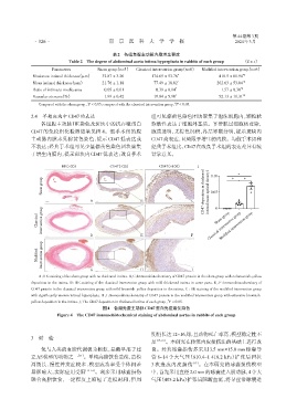
2.4 早期斑块中CD47的表达 组可见棕黄色染色团块聚集于泡沫细胞内,颗粒状
各组腹主动脉HE染色及斑块中的抗吞噬蛋白 弥散性表达于细胞外基质。平滑肌层细胞核蓝染,
CD47 的免疫组化检测结果见图 4。假手术组的腹 胞质透明,无棕色沉积,各层界限分明,提示斑块内
主动脉内膜未见棕黄色染色,提示 CD47 低表达或 CD47高表达,且局限于增生的内膜。与假手术组和
不表达;经典手术组可见少量棕黄色染色团块聚集 经典手术组比,CD47在改良手术组的表达差异有统
于增生内膜内,提示斑块内CD47低表达;改良手术 计学意义。
HE(×20) CD47(×20) CD47(×100) J *
Sham group deposition in thickened ( mean optical density) 0.10 * *
0.05
A B C CD47 intima 0
Modified intervention group
Sham group
Classical intervention group Classical intervention group
D E F
Modified intervention group
G H I
A:HE staining of the sham group with no thickened intima. B,C:Immunohistochemistry of CD47 protein in the sham group with no brownish⁃yellow
deposition in the intima. D:HE staining of the classical intervention group with mild thickened intima in some parts. E,F:Immunohistochemistry of
CD47 protein in the classical intervention group with mild brownish⁃yellow deposition in the intima. G:HE staining of the modified intervention group
with significantly uneven intimal hyperplasia. H,I:Immunohistochemistry of CD47 protein in the modified intervention group with extensive brownish⁃
*
yellow deposition in the intima. J:The CD47 deposition in thickened intima of each group,P < 0.05.
图4 各组兔腹主动脉CD47蛋白免疫组化染色
Figure 4 The CD47 immunohistochemical staining of abdominal aortas in rabbits of each group
期仍长达12~16周,且动物死亡率高,模型稳定性不
3 讨 论
足 [15-16] 。本研究在传统内皮损伤法的基础上进行改
兔与人类的血脂代谢极为相似,是最早用于建 良。经典球囊损伤多采用 3.5 mm×15.0 mm 球囊导
立AS模型的动物之一 [11] 。单纯高脂饮食造模,造模 管 8~14 个大气压(810.4~1 418.2 kPa)扩张后回拉
周期长、慢性并发症较多、模型成功率受个体间差 3 次造成内皮损伤 [17] 。在本研究的球囊损伤模型
异影响大,实际应用受限 [12-14] 。现多采用球囊损伤 中,首先采用直径2.0 mm的球囊进入股动脉,4个大
联合高脂饮食,一定程度上缩短了造模时间,但周 气压(405.2 kPa)扩张后阻断血流,将导丝沿球囊送