


Page 80 - 南京医科大学自然版
P. 80
第44卷第9期
               ·1252 ·                           南 京    医 科 大 学 学         报                        2024年9月


                                                      表1 磁共振扫描参数
                                           Table 1 Magnetic resonance scanning parameters

                                                               2D                               3D CS⁃SPACE
                     Parameter             Axial             Coronal            Sagittal          Coronal
                                       PdWI     T1WI     PdWI     T1WI      PdWI     T1WI     PdWI      T1WI
                TR(ms)                3 050.0   512.0   3 450.0   470.0    2 790.0   470.0    1 000     400
                TE(ms)                0 039.0   010.0   0 032.0   012.0    0 026.0   012.0    0 022     021
                FOV(mm)              160×160   160×160  180×180  180×180   180×180  180×180  220×220  220×220
                Thickness(mm)         0 003.0   003.0   0 003.0   003.0    0 003.0   003.0    0 001     001
                Interval(mm)          0 000.6   000.6   0 000.9   000.9    0 000.6   000.6    0 000     000
                Matrix(mm)           320×256   320×256  320×240  320×240   384×512  384×512  224×224  224×224
                Time(s)               0 106.0   140.0   0 146.0   110.0    0 140.0   110.0    0 234     217
                Accelerated factor(PI/CS)  0 002.0  002.0  0 002.0  002.0  0 002.0   002.0    0 003     003
                 Conventional 2D sequences were accelerated through parallel acquisition,whereas 3D CS⁃SPACE sequences employed compressed sensing for ac⁃
              celeration. PI:parallel imaging;CS:compressed sensing.

                                                                韧带(inferior acromioclavicular ligament,iACL)、斜方
                                                                韧带(trapezoid ligament,TL)、锥状韧带(conoid liga⁃
                                                                ment,CL)进行诊断评估,并给出诊断信心评分。韧
                                                                带损伤采用4分法进行评估(图2)。评估标准如下:
                                                                1,正常;2,损伤肿胀;3,部分撕裂;4,完全撕裂。诊
                                                                断信心评级同样采用4分法,其评级标准为:1,信心
                                                                欠佳;2,信心一般;3,信心较足;4,信心充足。将常
                                   A                       B
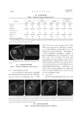
                                                                规 2D 图像和 3D CS⁃SPACE 图像分别交由上述 3 位
                 A:T1WI image of conventional 2D image. B:T1WI image of 3D CS
                                                                医师进行诊断评估并得到各自的评分结果,然后由
              ⁃SPACE.
                          图1 感兴趣区域勾画示意图                         3 位 医 师 共 同 商 量 决 定 得 到 2D 图 像 和 3D CS ⁃
                 Figure 1 Diagram of outlining the region of interest  SPACE 图像的诊断评估结果。上述医师进行个人
                                                                评估时,均先对常规 2D 图像进行评估,间隔 2 周后
              1.2.3 肩锁关节损伤的主观评价                                 再对 3D CS⁃SPACE 图像进行评估,评估时允许对
                  由3位分别具有3年、5年和10年以上肌骨影像                        3D CS⁃SPACE图像进行MPR。
              阅片经验的放射科医师对肩锁关节的上肩锁韧带                             1.3  统计学方法
             (superior acromioclavicular ligament,sACL)、下肩锁          使用SPSS 24.0统计软件进行分析,符合正态分


                                    A                       B                        C                       D
                 A:Grade 1(normal):the trapezoid ligament and conoid ligament indicated by arrows run continuously with normal signal. B:Grade 2(injury and
              swelling):ligament indicated by arrows runs continuously,with slightly elevated signal intensity in and around it. C:Grade 3(partial tear):the fiber bun⁃
              dles of the trapezoid ligament indicated by the arrow are still continuous. D:Grade 4(complete tear):the normal course of the trapezoid ligament indicated
              by the arrow is not visible,and the broken end is locally visible.
                                                    图2  韧带损伤分级示意图
                                      Figure 2  Schematic diagram of ligament injury classification

