


Page 108 - 南京医科大学自然版
P. 108
第45卷第2期
               ·248 ·                            南 京    医 科 大 学 学         报                        2025年2月


              A                         B
                                          Characteristies       HR  Lower 95%CI                 Upper 95%CI  P
                           ***            Male                 0.513   0.071                       3.722  <0.001
                                                                                                           0.509
                expression of SMC1A gene  7 6 5  Ajcc_pathoiogic_stageStage Ⅱ  1.859  1.003       26.912  <0.001
                 8
                                                                       1.022
                                                                                                   1.050
                                          Age_at_index
                                                               1.036
                                                                                                   3.446
                                                                                                           0.049
                                                                       1.401
                                                                                                           0.005
                                          Ajcc_pathoiogic_stageStage Ⅲ
                                                               3.111
                                                                                                   0.908
                                          Ajcc_pathoiogic_stageStage Ⅳ 11.038
                                                                       4.527
                                                                                                   2.133
                                                                                                           0.233
                                                               1.332
                                                                       0.832
                                          Ajcc_pathoiogic_nN1
                 4
                                          Ajcc_pathoiogic_nN2
                                                                                                           0.468
                                                                                                   3.016
                                          Ajcc_pathoiogic_nN3
                                                                       0.602
                                                               1.348
                 3
                                          Ajcc_pathoiogic_nNX  1.324   0.618                       2.840   0.470
                                                                       1.321
                                                               3.533
                                                                                                           0.012
                                                                                                   9.448
                      Normal  Turmor
                     (n=112) (n=1 093)    SMC1A                1.257   1.017                       1.554   0.035
                                                                            1     Hazard ratios
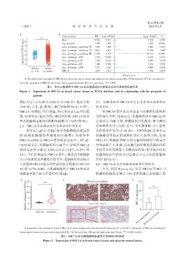
                 A:The differential expression of SMC1A gene in breast cancer tissues and normal breast tissues analyzed by TCGA database. B:The correlation be⁃
                                                                      ***
              tween the expression of SMC1A and breast cancer prognosis analyzed by Cox regression. P < 0.001.
                                图1 TCGA数据库中SMC1A在乳腺癌组织中的表达及其与患者预后的关系
              Figure 1  Expression of SMC1A in breast cancer tissues in TCGA database and its relationship with the prognosis of
                       patients
              预后不良),以年龄(0=≤60 岁,1=>60 岁)、临床分期                   2.3  乳腺癌组织SMC1A表达与患者临床病理特征
             (0=0期,1=Ⅱ、Ⅲ、Ⅳ期)、淋巴结转移(0=无,1=有)、                    的相关性
              SMC1A(实测值)为自变量,纳入多因素 Cox 回归模                           对 SMC1A 蛋白表达与患者不同临床特征的相
              型,结果显示:临床分期、淋巴结转移、SMC1A 表达                        关性进行分析,结果显示,乳腺癌组织中 SMC1A 蛋
              量是乳腺癌患者预后的影响因素(P < 0.05,图1B)。                     白表达与 TNM 分期、肿瘤组织分化程度、是否淋巴
              2.2  SMC1A在乳腺癌组织及癌旁组织的表达                          结转移有关(P < 0.05,表3),与患者年龄、分子亚型
                  采用 RT⁃qPCR 及 IHC 法分别检测临床收集到                   及肿瘤直径无关(P >0. 05)。同时将 IHC 总评分与
              的 48 例乳腺癌患者癌组织及癌旁正常组织中                            乳腺癌患者临床病理特征进行关联性分析,发现
              SMC1A mRNA 及 SMC1A 蛋白的表达水平。RT⁃qP⁃                 SMC1A 在乳腺癌 TNM 分期为Ⅲ期的组织中的总评
              CR 结果显示,乳腺癌组织与癌旁正常组织 SMC1A                        分明显高于Ⅰ/Ⅱ期,在有淋巴结转移的乳腺癌组织
              相对表达量分别为 4.204 ±2.145、1.012 ± 0.259(图             中的总评分显著高于无淋巴结转移,在中低分化的
              2A)。IHC结果显示,SMC1A蛋白主要表达于细胞核                       乳腺癌组织中的总评分显著高于高分化的乳腺癌
              内,以棕黄色及黄褐色着色为主,乳腺癌组织及癌旁                           组织(图3A~C)。
              正常组织SMC1A蛋白阳性表达率分别是87.5%(42/                      2.4  SMC1A表达对乳腺癌细胞凋亡的影响
              48)和16.7%(8/48),且乳腺癌组织中SMC1A的表达                        首先,利用 Western blot 鉴定 SMC1A 在 MCF⁃7
              强度显著高于癌旁正常组织(图2B)。                                乳腺癌细胞系及 MCF⁃10A 正常乳腺上皮细胞中的
               A   20         ***         B                Breast cancer tissues        C    15      ***
                  Relative expression of SMC1A  10 5 0      Para⁃tumor tissues              SMC1A staining score  10 5
                   15


                   -5
                        Adjacent Carcinoma                                                    0
                                                                                           Para⁃tumor tissues
                                                                                                Breast cancer tissues


                 A:Detection of the expression level of SMC1A in cancer tissues and adjacent normal tissues by RT⁃qPCR. B,C:Detection of SMC1A expression in
              breast cancer tissues and para⁃tumor tissues by IHC. The left scale bar:100 μm;the right scale bar:50 μm.  *** P < 0.001(n=48).
                                         图2 SMC1A在乳腺癌组织及癌旁正常组织中的表达
                              Figure 2  Expression of SMC1A in breast cancer tissues and adjacent normal tissues

