


Page 109 - 南京医科大学自然版
P. 109
第45卷第2期          丁   峰,喻夏飞,朱妍慧,等. SMC1A在乳腺癌中的表达及对乳腺癌细胞凋亡的影响[J].
                  2025年2月                     南京医科大学学报(自然科学版),2025,45(2):245-252                        ·249 ·


                                        表3 乳腺癌组织中SMC1A表达与患者临床病理特征的关系
                   Table 3 Relationship between SMC1A expression in breast cancer tissues and clinicopathological features of patients
                                                                                                        [n(%)]
                      Clinicopathological parameter     n            Positive          Negative          P
                    Age                                                                                 0.738
                      <60 years                        29           25(86.2)           4(13.8)
                      ≥60 years                        19           17(89.5)           2(10.5)
                    Maximum diameter of tumors                                                          0.827
                      <2.5 cm                          26           23(88.5)           3(11.5)
                      ≥2.5 cm                          22           19(86.4)           3(13.6)
                    Histological grading                                                                0.043
                      Low⁃moderate differentiation     30           24(80.0)           6(20.0)
                      High differentiation             18           18(100.0)          0(0)
                    Molecular types                                                                     0.857
                      Lumina A                         18           15(83.3)           3(16.7)
                      Lumina B                         15           13(86.7)           2(13.3)
                      Overexpression of HER⁃2          13           12(92.3)           1(7.7)
                      Three negative                    2           02(100.0)          0(0)
                    Stage of TNM                                                                        0.027
                      Stage Ⅰ/Ⅱ                        28           22(78.6)           6(21.4)
                      Stage Ⅲ                          20           20(100.0)          0(0)
                    Lymph node metastasis                                                               0.012
                      Existent                         23           23(100.0)          0(0)
                      Non⁃existent                     25           19(76.0)           6(24.0)


                         A                           B                            C
                             15       **                 15      ***                  10 8     ***
                            SMC1A staining score  10 5  SMC1A staining score  10 5   SMC1A staining score  6 4


                             0                            0                            2 0
                                (n=28)  (n=20)              (n=25)  (n=23)                (n=18)  (n=30)
                                                                                             Low⁃moderate
                             Ⅰ+Ⅱ     Ⅲ                     N0   N1+2                High differentiation
                                                                                          differentiation


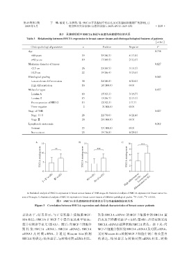
                   A:Statistical analysis of SMC1A expression in breast cancer tissues of TNM stages. B:Statistical analysis of SMC1A expression in breast cancer tis⁃
                                                                                          **
                                                                                                 ***
                sues of N stages. C:Statistical analysis of SMC1A expression in breast cancer tissues of different pathological grades. P < 0.01, P < 0.001.
                                       图3  SMC1A在乳腺癌组织中的表达水平与患者临床特征的关系
                        Figure 3  Correlation between SMC1A expression and clinical characteristics of breast cancer patients


                表达水平,结果显示,与正常乳腺上皮细胞 MCF⁃                          转染 SMC1A siRNA1 的 MCF⁃7 细胞中的 SMC1A 蛋
                10A 相比,SMC1A 在 MCF⁃7 中蛋白表达水平较高,                   白表达下降最明显(P < 0.05,图4B),后续实验采用
                差异有统计学意义(图4A)。随后,向MCF⁃7细胞分                        SMC1A siRNA1 敲降细胞 SMC1A 表达。接下来,向

                别 转 染 SMC1A siRNA1、SMC1A siRNA2、SMC1A             MCF⁃7细胞分别转染SMC1A siRNA1及对照siRNA,
                siRNA3 及 对 照 siRNA,并 通 过 Western blot 检 测         采用 Western blot 检测 MCF⁃7 细胞中凋亡相关蛋白
                SMC1A的表达;结果显示,与转染对照siRNA相比,                       的表达,结果显示与转染对照 siRNA 相比,转染

