


Page 110 - 南京医科大学自然版
P. 110
第46卷第10期
               ·1540 ·                           南 京    医 科 大 学 学         报                        2026年10月


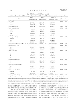
                                             表1 不同组胃癌患者的临床和病理特征比较
                  Table 1 Comparison of clinical and pathological characteristics among different groups of gastric cancer patients

                                                  HER2 absent      HER2 low        HER2 high
                                                                                                   2
                           Variables                                                              χ /H     P
                                                expression(n=35) expression(n=46)  expression(n=29)
               Sex[n(%)]                                                                          1.490   0.475
                 Male                             23(65.71)00   0.32(69.57)    0.023(79.31)
                 Female                           12(34.29)00   0.14(30.43)    0.006(20.69)
               Age[year,M(P25,P75)]               66(57,74)0    0.67(56,72)    0.068(66,76)       2.177   0.337
               Tumor location[n(%)]                                                               5.328   0.506
                 Cardia                            7(20.00)0    0.12(26.09)    0.008(27.59)
                 Fundus                            3(8.57)00    0.01(2.17)     0.008(27.59)
                 Body                             15(42.86)00   0.13(28.26)    0.011(37.93)
                 Antrum                           10(28.57)00   0.20(43.48)    0.002(6.90)
               Lauren type[n(%)]                                                                  8.999   0.061
                 Intestinal                        7(20.00)0    0.18(39.13)    0.015(51.72)
                 Mixed                            14(40.00)00   0.17(36.96)    0.010(34.48)
                 Diffuse                          14(40.00)00   0.11(23.91)    0.004(13.79)
               Maximum tumor diameter[cm,M(P25,P75)]  6.26(3.50,7.59)  5.50(3.39,7.00)  06.50(4.55,9.00)  4.145  0.126
               Differentiation[n(%)]                                                              8.138   0.017
                 Poor                             31(88.57)00   0.30(65.22)    0.017(58.62)
                 Moderate                          4(11.43)0    0.16(34.78) a  0.012(41.38) a
               AJCC stage[n(%)]                                                                   8.456   0.172
                 Ⅰ                                 2(5.71)00    0.02(4.35)     0.000(0.00)
                 Ⅱ                                 4(11.43)0    0.07(15.22)    0.001(3.45)
                 Ⅲ                                15(42.86)00   0.15(32.61)    0.007(24.14)
                 Ⅳ                                14(40.00)00   0.22(47.83)    0.021(72.41)
               Distant metastasis[n(%)]                                                           8.945   0.011
                 Yes                              14(40.00)00   0.22(47.83)    0.022(75.86)
                 No                               21(60.00)00   0.24(52.17)    0.007(24.14) ab
               Signet ring component[n(%)]                                                       15.955  <0.001
                 Yes                              17(48.57)00   0.13(28.26)    0.001(3.45) ab
                 No                               18(51.43)00   0.33(71.74)    0.028(96.55) ab
               CEA categorical[n(%)]                                                             44.444  <0.001
                 ≥5 ng/mL                          6(17.14)0    0.27(58.70) a  0.029(100.00) ab
                 <5 ng/mL                         29(82.86)00   0.19(41.30)    00.00(0) ab
               CEA[μg/L,M(P25,P75)]              2.17(1.37,3.89)  7.07(2.83,18.22) a  30.73(19.63,82.90) ab  52.206  <0.001

                 The serum CEA detection utilizes the standardized testing platform of our hospital’s clinical laboratory,with a cutoff value of 5 ng/mL(the recom⁃
              mended upper limit of normal reference for the reagent kit,which is the commonly used threshold in domestic clinical practice).Comparisons of age,le⁃
                                                                                                 2
              sion length and continuous serum CEA levels among the three groups were performed using the Kruskal⁃Wallis H test,while the χ test was adopted for
              comparisons of the remaining categorical variables. Post⁃hoc pairwise comparisons were performed using the Dunn test with Bonferroni correction ap⁃
              plied. a:compared to the HER2 absent expression group,P < 0.016 7;b:compared to the HER2 low expression group,P < 0.016 7.
              低表达人群基数庞大,HER2 低表达状态的精准识                         建预测胃癌HER2表达模型 等。 F⁃FDG PET/CT以
                                                                                        [22]
                                                                                              18
              别是该人群能否从抗HER2靶向治疗中受益的前提。                          其应用广泛、无创、可重复、可半定量评估肿瘤代谢活
                  目前,手术或活检组织的IHC联合FISH检测是                       性及全身一站式成像等优点,已在多项研究中被证实
                                                                                                  [12]
              评估 HER2 表达状态的“金标准”,但该方法存在有                        其代谢参数可预测胃癌HER2表达阳性 。Park等                   [23]
              创性等固有局限,近年来越来越多的研究转向探索                            研究发现,HER2 阳性组的 PET 代谢参数 SUVmax
              非侵入性、智能化替代方案,包括应用深度学习模型                           明显高于阴性组。本研究将 HER2 表达状态分为
                              [21]
              分析胃癌病理切片 、以及基于能谱CT定量参数构                           3 组,分析发现 MTV 80%、TLG 80%、SUVmax 以及 HI 在

