
Page 50 - 《南京医科大学学报》自然科学版2026年第2期
P. 50
第46卷第2期
·206 · 南 京 医 科 大 学 学 报 2026年2月
A Sham TBI 3 Days B TBI
(×10) (×40) (×10) (×40) Sham 3 h 1 d 3 d 7 d 14 d
Dixdc1 77 kDa
GFAP 50 kDa
α⁃Tubulin 55 kDa
TBI 3 hours TBI 7 Days
(×10) (×40) (×10) (×40)
A C
1.5 **
***
Dixdc1/α⁃Tubulin 1.0
TBI 1 Day TBI 14 Days
(×10) (×40) (×10) (×40) 0.5
0
Sham 3 h 1 d 3 d 7 d 14 d
TBI
(×100) (×200)
D E F
*** 25
4 3 *** *** (mm 2 ) ***
GFAP/α⁃Tubulin 2 1 Sham positive area 20 5
15
10
0 Dixdc1 0
Sham 3 h 1 d 3 d 7 d 14 d 3 h Sham TBI 3 h
TBI
TBI
G (×100) (×200) H
(mm 2 ) 25 ***
GFAP positive area 20 5
Sham 15
10
3 h 0 Sham TBI 3 h
TBI
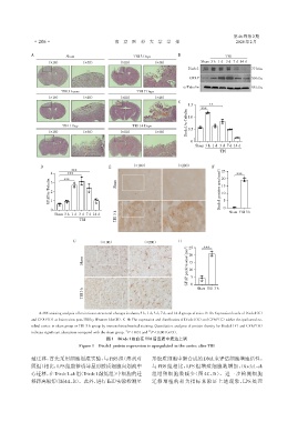
A:HE staining analysis of brain tissue structural changes in sham,3 h,1 d,3 d,7 d,and 14 d groups of mice. B-D:Expression levels of Dixdc1(C)
and GFAP(D)at lesion sites post⁃TBI by Western blot(B). E-H:The expression and distribution of Dixdc1(E)and GFAP(G)within the ipsilateral ce⁃
rebral cortex in sham group or TBI 3 h group by immunohistochemical staining. Quantitative analyses of protein density for Dixdc1(F)and GFAP(H)
**
indicate significant alterations compared with the sham group. P < 0.01 and *** P < 0.001(n=3).
图1 Dixdc1蛋白在TBI后皮质中表达上调
Figure 1 Dixdc1 protein expression is upregulated in the cortex after TBI
殖迁移,首先采用细胞划痕实验,与 PBS 组(溶剂对 形胶质细胞中新合成的DNA来评估细胞增殖活性。
照组)相比,LPS组能够诱导星形胶质细胞向划痕中 与 PBS 组相比,LPS 组增殖细胞数增加,Dixdc1⁃sh
心迁移,在Dixdc1⁃sh组(Dixdc1敲低组)中细胞的迁 组增殖细胞数减少(图 4C、D)。进一步检测细胞
移距离缩短(图4A、B)。此外,进行EdU实验检测星 迁移增殖的相关指标来验证上述现象,LPS 处理