
Page 51 - 《南京医科大学学报》自然科学版2026年第2期
P. 51
第46卷第2期 汤莉巧,段程伟,陈伟观,等. 下调Dixdc1表达抑制创伤性脑损伤后星形胶质细胞极化[J].
2026年2月 南京医科大学学报(自然科学版),2026,46(2):202-212 ·207 ·
A (×200) (×400) C (×200) (×400)
Lesion core Lesion core
Sham Iba1
Lesion core
Lesion core
3 h NenN
TBI
(Unit) 200 Dixdc1 (Unit) 250 Dixdc1 (Unit) 250 Iba1 (Unit) 200 NeuN
B Sham TBI 3 h D
intensity 150 GFAP intensity 200 GFAP intensity 200 Dixdc1 intensity 150 Dixdc1
150
150
100
100
100
100
Fluorescent 50 0 Fluorescent 50 0 Fluorescent 50 0 Fluorescent 50 0
40
80 100
60
200
400
0
Distance(μm)
Distance(μm)
Distance(μm) 600 0 100 200 300 400 0 50 100 150 200 250 0 20 Distance(μm)
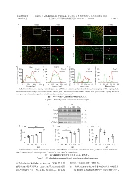
A,B:Immunofluorescence staining of Dixdc1(green)and GFAP(red)within the ipsilateral cerebral cortex in sham group or TBI 3 h group. C,D:
Immunofluorescence staining of Dixdc1(red)and Iba1/NeuN(green)within the ipsilateral cerebral cortex in sham group or TBI 3 h group. The lesion
core region was delineated using a white dashed line and annotated as“Lesion core”.
图2 Dixdc1蛋白与星形胶质细胞存在共定位
Figure 2 Dixdc1 protein co⁃localizes with astrocytes
A LPS
0 h 4 h 8 h 12 h 24 h
Dixdc1 77 kDa
GFAP 50 kDa
PCNA 36 kDa
α⁃Tubulin 55 kDa
B C * D *
3.0 *** 2.5 2.2
** 2.1
Dixdc1/α⁃Tubulin 2.0 * GFAP/α⁃Tubulin 1.5 PCNA/α⁃Tubulin 2.0
***
2.0
**
1.9
1.0
1.0
1.7
0 0.5 0 1.8
0 h 4 h 8 h 12 h 24 h 0 h 4 h 8 h 12 h 24 h 0 h 4 h 8 h 12 h 24 h
LPS LPS LPS
A:Western blot detection of protein levels of Dixdc1,GFAP and PCNA in an activated astrocyte model. B-D:Quantitative analysis of Dixdc1(B),
*
**
GAFP(C)and PCNA(D)protein expressions. P < 0.05,P < 0.01,and *** P < 0.001(n=3).
图3 LPS刺激促进星形胶质细胞中Dixdc1蛋白表达
Figure 3 LPS stimulation promotes Dixdc1 protein expression in astrocytes
后 E⁃Cadherin、N⁃Cadherin、Vimentin、PCNA 的蛋白 制星形胶质细胞增殖迁移能力。
表达较PBS处理组增加,Dixdc1⁃sh组Dixdc1敲低抑 2.5 敲低Dixdc1抑制LPS诱导的星形胶质细胞周期
制 LPS 的诱导作用(图 4E~J)。提示 Dixdc1 敲低抑 细胞周期是检测细胞增殖的重要检测指标 [18] ,