


Page 28 - 南京医科大学自然版
P. 28
第46卷第8期
               ·1134 ·                           南 京    医 科 大 学 学         报                        2026年8月


              肿块和 CPH⁃I 联合诊断比 O⁃RADS 分类单独诊断                     简单法则单独诊断附件肿块的 AUC 更高,但差异
              附件肿块的 AUC 更高(Z=2.285,P=0.022)。IOTA                无统计学意义(Z=0.580,P=0.562)。典型病例见
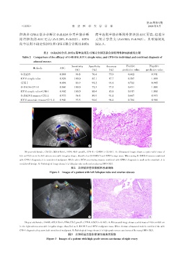
              简单法则不确定性肿块和 CPH⁃I 联合诊断比 IOTA                      图 2、3。

                              表2 O⁃RADS分类、IOTA简单法则及CPH⁃I分别及联合诊断附件肿块的效能比较
                Table 2 Comparison of the efficacy of O⁃RADS,IOTA simple rules,and CPH⁃I in individual and combined diagnosis of
                       adnexal masses
                                                 Sensitivity  Specificity  Accuracy  Positive       Negative
                       Methods           AUC
                                                  (%)        (%)         (%)      predictive value  predictive value
               O⁃RADS                    0.866    096.8       76.4       79.9         0.462           0.991
               IOTA simple rules         0.926    100.0       85.1       87.7         0.585           1.000
               CPH⁃I                     0.886    083.9       93.2       91.6         0.722           0.965
               O⁃RADS+CPH⁃I              0.861    100.0       72.3       77.1         0.431           1.000
               IOTA simple rules+CPH⁃I   0.902    100.0       80.4       83.8         0.517           1.000
               O⁃RADS 4 masses+CPH⁃I     0.933    096.8       89.9       91.1         0.667           0.993
               IOTA uncertain masses+CPH⁃I  0.941  093.5      94.6       94.4         0.784           0.986


                                                            A                                       B

                 38⁃year⁃old female,CA125:205.6 U/mL,HE4:46.9 pmol/L,CPH⁃I:-2.489(<-2.165). A:Ultrasound image shows a cystic⁃solid mass of
              6.6 cm×5.0 cm in the left adnexa area with irregular shape,classified as O⁃RADS 5 and IOTA benign mass. When using O⁃RADS 4 masses combined
              with CPH⁃I diagnosis,it is considered malignant. While when IOTA uncertainty masses combined with CPH⁃I diagnosis is used as the standard,it is
              considered benign. B:Pathological image shows it is fallopian tube and ovarian abscess(HE×100).
                                                图2 左侧输卵管卵巢脓肿患者图像
                                  Figure 2 Images of a patient with left fallopian tube and ovarian abscess


                                                            A                                       B
                 50⁃year⁃old female,CA125:455.9 U/mL,HE4:578.7 pmol/L,CPH⁃I:2.387(>-2.165). A:Ultrasound image shows a solid mass of 12.6 cm×8.8 cm
              in the right adnexa area with irregular shape,classified as O⁃RADS 5 and IOTA malignant mass. When the two ultrasound models combined the with
              CPH⁃I diagnosis,they were both considered malignant. B:Pathological image shows it is high⁃grade serous carcinoma of the ovary(HE×100).
                                              图3 右侧卵巢高级别浆液性癌患者图像
                                Figure 3  Images of a patient with high⁃grade serous carcinoma of right ovary

