


Page 42 - 南京医科大学自然版
P. 42
第46卷第8期
               ·1148 ·                           南 京    医 科 大 学 学         报                        2026年8月


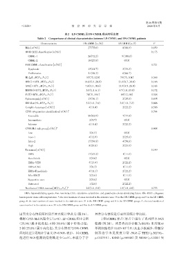
                                            表2 LP⁃CMML及DN⁃CMML临床特征比较
                          Table 2 Comparisons of clinical characteristics between LP⁃CMML and DN⁃CMML patients

                            Characteristics              DN⁃CMML(n=36)          LP⁃CMML(n=9)           P
                  Male[n(%)]                                27(75.0)               8(88.9)            0.659
                  WHO 2022 classification[n(%)]                                                       0.173
                    CMML⁃1                                  26(72.2)              9(100.0)
                    CMML⁃2                                  10(27.8)                0(0)
                  FAB CMML classification[n(%)]                                                       0.721
                    Dysplastic                              15(41.7)               3(33.3)
                    Proliferative                           21(58.3)               6(66.7)
                  Hb[g/L,M(P25,P75 )]                      95(75,120)            79(73,104)           0.348
                  WBC[×10 /L,M(P25,P75 )]                 14.6(5.9,20.8)        21.0(8.7,38.6)        0.146
                         9
                  ANC[×10 /L,M(P25,P75 )]                 7.4(3.3,10.6)         13.9(4.5,26.0)        0.163
                         9
                  MONO[×10 /L,M(P25,P75 )]                 3.0(1.1,6.1)          4.7(1.8,13.8)        0.172
                           9
                  PLT[×10 /L,M(P25,P75 )]                  74(31,161)             68(12,94)           0.348
                         9
                  Splenomegaly[n(%)]                        13(36.1)               2(25.0)            0.695
                  BM blast[%,M(P25,P75 )]                  5.2(1.6,7.6)          3.6(1.8,7.2)         0.606
                  Complex karyotype[n(%)]                   4(11.4)                2(22.2)            0.586
                  CPSS cytogenetics classification[n(%)] *                                            0.744
                    Favorable                               29(82.9)               7(77.8)
                    Intermediate                             2(5.7)                 0(0)
                    Adverse                                 4(11.4)                2(22.2)
                  CPSS⁃Mol risk group[n(%)] #                                                         0.905
                    Low                                      2(6.5)                 0(0)
                    Inter⁃1                                 4(12.9)                2(25.0)
                    Inter⁃2                                 17(54.8)               4(50.0)
                    High                                    8(25.8)                2(25.0)
                  Treatment[n(%)]                                                                     0.149
                    HMA                                     17(47.2)               1(11.1)
                    Ruxolitinib                              2(5.6)                 0(0)
                    HMA+VEN                                 5(13.9)                2(22.2)
                    HMA+CAG                                  3(8.3)                1(11.1)
                    HMA+Ruxolitinib                         4(11.1)                2(22.2)
                    Allo⁃HSCT                                2(5.6)                1(11.1)
                    Supportive care                          2(5.6)                 0(0)
                    Untreated                                1(2.8)                2(22.2)
                  Numbers of HMA courses[M(P25,P75 )] △    2.0(1.0,5.0)          1.5(1.0,4.0)         0.733
                 HMA:hypomethylating agents;Ven:venclexta;CAG:cytarabine,aclarubicin,and granulocyte colony⁃stimulating factor;Allo⁃HSCT:allogeneic
              hematopoietic stem cell transplantation. *:the total numbers of cases involved in the statistics were 35 in the DN⁃CMML group and 9 in the LP⁃CMML
              group;#:the total numbers of cases involved in the statistics were 31 in the DN⁃CMML group and 8 in the LP⁃CMML group;△:the total numbers of
              cases involved in the statistics were 31 in the DN⁃CMML group and 8 in the LP⁃CMML group.

              因突变分布情况两组间差异无统计学意义(图2A)。                          异常分布情况进行亚组间统计学比较。
              根据 CPSS⁃Mol 风险分层(n=8),LP⁃CMML 组中 2 例                   1 例 tCMML 患者(患者 7)进行了系列样本 NGS
             (25.0%)属于低危组,4 例(50.0%)属于中危⁃2 组,                   检测(图 2B)。该患者初诊诊断为 BLPD,血常规示
                                                                                     9
              2 例(25.0%)属于高危组,其分布情况与 DN⁃CMML                    单核细胞增多(0.65×10 个/L)及血小板减少,骨髓穿
              组相比差异无统计学意义(P=0.905,表2)。因tCMML                    刺形态学未见发育异常,NGS 示 TET2(p.N275fs、
              组进行 NGS 检测的病例数过少(n=3),未就分子学                       p.C1273Y)、EZH2(p.H694R)及 NRAS(p.G12D)突

