


Page 122 - 南京医科大学自然版
P. 122
第46卷第9期
               ·1392 ·                           南 京    医 科 大 学 学         报                        2026年9月


                        Imaging and             Interface           Proximity labeling        Functional
                        visualization           enrichment          and spatial profiling     validation

                  TEM    CLEM    Cryo⁃ET                             Split⁃TurbolD
                                                 Cell
                                                               APEX2
                                                 homogenization              Contact⁃ID  Fatty acid
                                                                                                   β⁃oxidation
                                                                                          transfer
                                                                                                     ROS
                                                 Density⁃gradient
                 Super⁃resolution  Live⁃cell     flotation                             ATP/OXPHOS
                   microscopy  imaging
                                                                                         mtDNA     Cell injury
                                                                                         release
                                              LD⁃associated
                                              mitochondria/PDM     Spatial proteomics
                                                                                          Inflammation/fibrosis
                  Visualize contact architecture  Enrichment for proteomics,  Define local molecular identity  Link contact remodeling to
                       and dynamics      lipidomics,and bioenergetics  of contact sites   functional outcomes


                                            Morphology          Composition       Function


                                                         Multi⁃layer validation
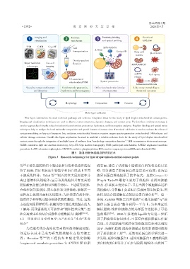
                 This figure summarizes the main technical pathways and evidence integration ideas for the study of lipid droplet mitochondrial contact points.
              Imaging and visualization techniques are used to observe contact structures,dynamic changes,and contact areas. The interface enrichment strategy is
              used to separate lipid droplet related mitochondria and conduct proteomics,lipidomics,and bioenergetics analyses. Neighbor labeling and spatial omics
              techniques help to analyze the local molecular composition and spatial features of contact sites. Functional validation is used to evaluate the effects of
              contact remodeling on fatty acid transport,beta oxidation,mitochondrial function,reactive oxygen species generation,mitochondrial DNA release,and
              cellular damage outcomes. Overall,the figure emphasizes the need to establish a reliable evidence chain for the study of lipid droplet mitochondrial
              contact points through the integration of multiple layers of evidence from“morphology composition function”. TEM:transmission electron microscopy;
              CLEM:correlative light and electron microscopy;Cryo⁃ET:Cryo electron tomography;PDM:peridroplet mitochondria;APEX2:engineered ascorbate
              peroxidase 2;ATP:adenosine triphosphate;OXPHOS:oxidative phosphorylation;ROS:reactive oxygen species;mtDNA:mitochondrial DNA.
                                                图3 脂滴⁃线粒体接触点的研究技术
                                 Figure 3 Research technology for lipid droplet mitochondrial contact points


                [43]
              等 在棕色脂肪组织中通过温和匀浆和低速浮选保                            滴表面,建立了活细胞中脂滴蛋白组的邻近标记策
              留了PDM,并证明其在生物能学和蛋白组成上不同                           略,显著提高了脂滴蛋白组鉴定的可信度,也为后
              于胞质线粒体。Talari 等       [16] 则首次在大鼠肝脏中分             续界面蛋白筛选提供了技术范式。虽然 Contact⁃ID
              离出脂滴相关线粒体,显示该类线粒体具有更高的                            和 split⁃TurboID 最初主要用于线粒体⁃内质网接触
              脂肪酸氧化能力和特征性酶学特征。上述研究说明,                           位点,但其核心优势在于:只有当两个细胞器在近距
              合理控制匀浆强度、离心条件和分层策略,能够在一                           离接触时,分裂酶才会重组并完成局部生物素化,因
              定程度上保留界面相关线粒体,为后续蛋白质组学、                           此特别适合膜接触位点的原位蛋白谱分析 。进一
                                                                                                      [45]
              脂质组学和呼吸功能分析提供物质基础。不过,这类                           步地,CsFiND 等新工具开始将“可视化接触”与“接
              方法的局限同样明显:机械剪切可能造成接触点的人                           触位点蛋白捕获”整合到同一平台上,为未来直接
              工解离,而残留黏附又可能带来“假阳性共富集”,因                          解析脂滴⁃线粒体接触点的局部蛋白组提供了可借
              此分离效果必须结合成像和功能数据共同解释                    [42-44] 。  鉴的路径    [46] 。2026 年报道的 LipoID 方法进一步拓

              4.3  邻近标记与空间组学:从“共定位”走向“界面                        展了脂滴邻近标记技术,可在原位捕获脂滴互作蛋
              组分”                                               白组,并识别脂滴与线粒体等细胞器互作相关调控
                  为克服传统分离纯化带来的污染和解离问题,                          因子,为解析脂滴⁃线粒体接触点的动态调控网络提
              邻近标记技术已成为研究膜接触位点的关键工                              供了新的技术工具 。近期邻近蛋白组学研究进一
                                                                                 [47]
              具。Bersuker 等   [44] 将工程抗坏血酸过氧化物酶                  步发现,延伸突触蛋白1、延伸突触蛋白2、囊泡相关膜
             (engineered ascorbate peroxidase 2,APEX2)靶向脂       蛋白相关蛋白B等分子可参与脂滴⁃线粒体⁃内质网三

