
Page 143 - 南京医科大学学报自然科学版
P. 143
第41卷第7期 张淬锋,李铭铭,冷迪雅,等. 牙源性上颌窦炎的锥形束CT影像学特征研究[J].
2021年7月 南京医科大学学报(自然科学版),2021,41(07):1069-1074 ·1073 ·
E
A B C D
F
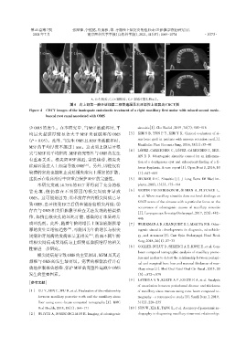
A、B:矢状位;C、D:冠状位;E、F:轴位(箭头所示)。
图4 右上颌第一磨牙近颊第二根管遗漏及右牙源性上颌窦炎CBCT图
Figure 4 CBCT images of the inadequate endodontic treatment of a right maxillary first molar with missed second mesio⁃
buccal root canal associated with OMS
少 OMS 的发生。在本研究中,当 MSF 被破坏时,平 sinusitis[J]. Clin Radiol,2019,74(7):503-516
均最大黏膜厚度显著大于 MSF 未被破坏的 OMS [3] KIM S B,YUN P Y,KIM Y K. Clinical evaluation of si⁃
(P < 0.05)。此外,当发生OMS且MSF未被破坏时, nus bone graft in patients with mucous retention cyst[J].
MSF 的平均厚度不超过 1 mm。这表明上颌后牙根 Maxillofac Plast Reconstr Surg,2016,38(1):35-40
[4] LÓPEZ⁃CARRICHES C,LÓPEZ⁃CARRICHES I,BRY⁃
尖与MSF的平均距离、MSF的完整性与OMS的发生
AN R B. Odontogenic sinusitis caused by an inflamma⁃
有重要关系。根尖离 MSF 越近,窦底越薄,根尖炎
tion of a dentigerous cyst and subsequent finding of a fi⁃
症越容易进入上颌窦导致OMS [27] 。另外,即使仅有 brous dysplasia. A case report[J]. Open Dent J,2016,10
较薄的窦底也能阻止或延缓炎症向上颌窦的扩散, (1):647-655
这提示在临床治疗中应重点保护MSF的完整性。 [5] BECKER D G. Sinusitis[J]. J Long Term Eff Med Im⁃
本研究发现 14.75%的 RCT 牙得到了充分的根 plants,2003,13(3):175-194
管充填,但仍存在不明原因的根尖周病并导致 [6] VESTIN FREDRIKSSON M,ÖHMAN A,FLYGARE L,
OMS。这可能是因为:①术前存在的根尖周病已导 et al. When maxillary sinusitis does not heal:findings on
CBCT scans of the sinuses with a particular focus on the
致 OMS,患牙接受 RCT 后仍未能治愈根尖周病;②
occurrence of odontogenic causes of maxillary sinusitis
存在与 OMS 相关但影像学检查无法发现的错误操
[J]. Laryngoscope Investig Otolaryngol,2017,2(6):442-
作,如挤出根尖孔的坏死牙髓、感染的牙本质碎片
446
或冲洗剂。此外,随着年龄的增长上颌窦底黏膜增 [7] WORKMAN A D,GRANQUIST E J,ADAPPA N D. Odon⁃
厚的发生呈增加趋势 [28] ,可能因为年龄增长与根尖 togenic sinusitis:developments in diagnosis,microbiolo⁃
周病和牙周病的发病率呈正相关 [29] ,然而不同年龄 gy,and treatment[J]. Curr Opin Otolaryngol Head Neck
组根尖周病或牙周病与上颌窦底黏膜增厚的相关 Surg,2018,26(1):27-33
性需进一步研究。 [8] GOLLER⁃BULUT D,SEKERCI A E,KÖSE E,et al. Cone
beam computed tomographic analysis of maxillary premo⁃
根尖周病是导致OMS的主要原因,MFM及其近
lars and molars to detect the relationship between periapi⁃
颊根与 OMS 的发生最密切。完善的根管治疗并有
cal and marginal bone loss and mucosal thickness of max⁃
效地控制根尖感染、保护MSF的完整性是减少OMS illary sinus[J]. Med Oral Patol Oral Cir Bucal,2015,20
发生的重要因素。 (5):e572-e579
[9] LATHIYA V N,KOLTE A P,KOLTE R A,et al. Analysis
[参考文献]
of association between periodontal disease and thickness
[1] GU Y,SUN C,WU D,et al. Evaluation of the relationship of maxillary sinus mucosa using cone beam computed to⁃
between maxillary posterior teeth and the maxillary sinus mography ⁃ a retrospective study[J]. Saudi Dent J,2019,
floor using cone ⁃ beam computed tomography[J]. BMC 31(2):228-235
Oral Health,2018,18(1):164-171 [10] SUN W,XIA K,TANG L,et al. Accuracy of panoramic ra⁃
[2] WHYTE A,BOEDDINGHAUS R. Imaging of odontogenic diography in diagnosing maxillary sinus⁃root relationship: