
Page 72 - 南京医科大学学报自然科学版
P. 72
第42卷第3期
·374 · 南 京 医 科 大 学 学 报 2022年3月
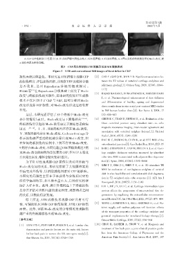
A B C D
A:CEP局灶性缺损(白色箭头);B:T1加权图像呈阴性表现;C:T2压脂图像;D:T2加权图像,B~D图示损伤的终板附近未见Modic改变,提
示损伤未累及椎体骨髓。
图3 CEP局灶性缺损UTE图像及常规MR图像表现
Figure 3 UTE and conventional MR images of focal defect in CEP
损伤面积具体量化。本研究在UTE图像上观察CEP [2] CAO Y,GUO Q W,WAN Y D. Significant association be⁃
的损伤程度、评估损伤面积,并观察VEP及椎体骨髓 tween the T2 values of vertebral cartilage endplates and
是 否 受 累 ,是 对 Rajasekaran 分 级 的 细 致 探 讨 。 pfirrmann grading[J]. Orthop Surg,2020,12(4):1164-
1172
Määttä等 [12] 在 Rajasekaran 分级基础上研究了 Modic
[3] RAJASEKARAN S,VENKATADASS K,NARESH BABU
改变与终板损伤的关联性,而本研究使用UTE成像
J,et al. Pharmacological enhancement of disc diffusion
技术不仅区分出了 CEP 与 VEP,且可以看到 Modic
and differentiation of healthy,ageing and degenerated
改变形成伴 VEP 损伤,对 Modic 改变形成过程有所 discs:results from in⁃vivo serial post⁃contrast MRI studies
补充。 in 365 human lumbar discs[J]. Eur Spine J,2008,17
最后,本研究还评估了 85 个终板中 Modic 改变 (5):626-643
存在的情况(表2)。Modic 改变与下腰痛相关 [13-14] , [4] CHEN K C,TRAN B,BISWAS R,et al. Evaluation of the
终板损伤后引起的 Modic改变也是下腰痛患者的病 Disco ⁃ vertebral junction using ultrashort time ⁃ to ⁃ echo
因之一 [15-16] 。Ⅰ、Ⅱ、Ⅲ级终板均不存在Modic改变, magnetic resonance imaging:inter⁃reader agreement and
association with vertebral endplate lesions[J]. Skeletal
Ⅴ、Ⅵ级终板均存在Modic改变,Cochran⁃Armitage分
Radiol,2016,45(9):1249-1256
析示损伤越严重的终板越容易出现 Modic 改变,但
[5] BAE W C,BISWAS R,CHEN K,et al. UTE MRI of the
在Ⅳ级终板损伤的病例中,7 例不存在 Modic 改变, osteochondral junction[J]. Curr Radiol Rep,2014,2(2):35
9 例存在Modic改变,本研究通过UTE图像发现这7例 [6] BERG⁃JOHANSEN B,HAN M,FIELDS A J,et al. Carti⁃
无Modic改变的病例均是仅累及CEP,但VEP未有或 lage endplate thickness variation measured by ultrashort
并未完全累及,椎体骨髓未受累(图3)。 echo⁃time MRI is associated with adjacent disc degenera⁃
为了针对性地观察 CEP 损伤以及对应终板与 tion[J]. Spine,2018,43(10):E592-E600
Modic 改变间的关系,本研究排除了其他腰椎实质 [7] KIM Y J,CHA J G,SHIN Y S,et al. 3D ultrashort TE
MRI for evaluation of cartilaginous endplate of cervical
性病变及外伤等,以消除其他因素对 CEP 的影响。
disk in vivo:feasibility and correlation with disk degenera⁃
本研究的局限性主要在于未获得与分级相对应的
tion in T2⁃weighted spin⁃echo sequence[J]. AJR Am J
组织学病理结果,并且样本量不大,后续研究将继
Roentgenol,2018,210(5):1131-1140
续扩大样本量。此外,将计算椎间盘上下终板损伤 [8] LIU L,HE J Y,LIU C,et al. Cartilage intermediate layer
总分并评估对应椎间盘的退变程度,观察两者之间 protein affects the progression of intervertebral disc de⁃
的关系,以期获得新发现。 generation by regulating the extracellular microenviron⁃
综上所述,UTE 成像技术观察 CEP 具有可行 ment(Review)[J]. Int J Mol Med,2021,47(2):475-484
性,可辅助临床评估 CEP 损伤程度,量化 CEP 损伤 [9] WONG J,SAMPSON S L,BELL⁃BRIONES H,et al. Nu⁃
面积。此外,评估 Modic 改变的分布情况发现损伤 trient supply and nucleus pulposus cell function:effects
of the transport properties of the cartilage endplate and
越严重的终板越容易出现Modic改变。
potential implications for intradiscal biologic therapy[J].
[参考文献] Osteoarthritis Cartilage,2019,27(6):956-964
[1] LIVSHITS G,POPHAM M,MALKIN I,et al. Lumbar disc [10] CHOU R,QASEEM A,SNOW V,et al. Diagnosis and
degeneration and genetic factors are the main risk factors treatment of low back pain:a joint clinical practice guide⁃
for low back pain in women:the UK twin spine study[J]. line from the American College of Physicians and the
Ann Rheum Dis,2011,70(10):1740-1745 American Pain Society[J]. Ann Intern Med,2007,147