Page 112 - 南京医科大学学报自然科学版
P. 112

第44卷第3期
               ·402 ·                          南   京 医 科       大 学      学 报                        2024年3月


                                                                                                       Skull
                                   Meninges structure                  CCL21
                                                                                                +
                                                                                            -
                                                                                       +
                                                                                    CD4 ,CD45RA ,CD27 ,CD69 +
                                                                                     IL⁃1
                                                                            CCR7
                              Cerebrum
               sCLN
                                                IL⁃6                                                  Dura mater
                                               TNF⁃α
                                                                                                      Arachnoid
                                                                                                        mater
                                                                                                       SLYM
                                                                   IL⁃4
                                                                  IL⁃13
                                   dCLN                      CD206
                                                                                                      Pia mater
                                                                                 RDNF
                                                                                 ICF⁃1
                                                                                 TGF⁃β
                                                                                                         Brai
                                                                                                      parcnchyma
                                                                 +
                                                    MLV       CD4 T cell  DC       Macrophage    Astrocyte
                                PROX1 +
                                                                 +
                                VEGFR3 +            Vein      CD8 T cell  ILC       MC           Microglia
                                LYVE1 +
                                CCL21 +
                        MLV                         Artery    NK cell     B cell   Monocyte      Neuron cell
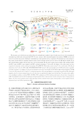
                 The dura mater can be divided into outer and inner layers,the arachnoid mater consists of a layer of closely connected outer cells,surrounding the
              subarachnoid space filled with cerebrospinal fluid(CSF). Recently,a structure called subarachoid lymphatic⁃like membrane(SLYM)was discovered
              between the arachnoid and pia mater,dividing the subarachnoid space into superficial and deep compartments. The innermost layer of the meninges is
              the pia mater,beneath which lies a membrane formed by closely connected astrocytes and their processes,known as the glia limitans. Together,the pia
              mater and glia limitans constitute the final barrier above the brain parenchyma. The dura mater contains numerous immune cells,including dendritic
              cells(DC),mast cells(MC),innate lymphoid cells(ILC),meningeal macrophages,T cells and B cells. Immune cells on the pia mater are fewer
              compared to the dura mater,mainly consisting of macrophages,DC and MC. The CSF primarily contains memory T cells,a small number of B cells,and
                                                         +
                                                              +
                                                     ⁃
                                              +
              mononuclear cells. T cells in the CSF include CD4 /CD45RA /CD27 /CD69 memory T cells expressing high levels of CCR7 and L⁃selectin,responsible
                                                                                                            +
              for immune surveillance. If no antigens are detected,they flow back into the deep cervical lymph nodes(dCLN)through meningeal lymphatics. CD4 αβ
              T cells in the CSF release IL⁃4,IL⁃13 and IL⁃17,exerting brain⁃protective effects and participating in social behavior by producing IFN⁃γ. Meningeal
              lymphatics,tube⁃like structures arranged along veins on the dura mater,are particularly developed around the skull base. They express classic markers
              under physiological conditions,including PROX1,VEGFR3,LYVE1,podoplanin and CCL21,lacking smooth muscle cells and valves. Comparatively
              smaller than lymphatics associated with the diaphragm,meningeal lymphatics form a relatively simple network covering fewer tissues. Carrying numerous
              immune cells,they connect cytokines related to brain parenchyma with the peripheral immune system,draining immune cells and cytokines from the
              subarachnoid space into dCLN and partially into sCLN.
                                               图1 脑膜结构和免疫功能相关示意图
                                         Figure 1 Structure and immune function of meninges


              泛,大脑灰质的轴突丢失也随之发生,脑膜炎症的                            型是 EAE 模型鼠,目前主要通过自身反应性 T 细胞
              严重性与 MS 病情严重程度和死亡率呈正相关。                           过继转移诱导被动性EAE模型鼠,或通过髓磷脂抗
              Lucchinetti 等 [34] 通过活体脑组织成像技术,发现脑                原免疫注射诱导主动性 EAE 模型鼠 。有研究在
                                                                                                 [5]
              膜炎症常常发生在 MS 早期,甚至可能早于白质病                          EAE 鼠的病理过程中观察到,有活性的 T 细胞进入
              变的出现。在MS病程中,大脑活性T细胞入侵中枢                           脑实质,而无活性的T细胞则进入CSF,经过软脑膜
              神经系统,诱导自我损伤性炎症反应发生,T细胞浸                           的筛选,T 细胞得以接触抗原,引起脑组织损伤,诱
              润最早在脑膜中被观察到,早于脑实质浸润以及临                            导 MS 的病理过程      [36] 。Furtado 等 [37] 发现,在 EAE 病
                                                         [35]
              床症状的出现,而在CSF中也发现了T细胞浸润 。                          理进程中,T细胞活化最早发生在颈淋巴结,手术切
              由此可见,脑膜炎症与MS的发病和进展紧密相关。                           除颈淋巴结可以改善EAE的病理情况,进一步证实
                  由于 MS 患者标本不易获取,最理想的研究模                        了EAE的发生与脑膜T细胞活化相关。Louveau 等                 [18]
   107   108   109   110   111   112   113   114   115   116   117