Page 41 - 南京医科大学自然版
P. 41

第44卷第4期            钱鑫娜,朱文卿,邱 憬. 牛磺鹅去氧胆酸对兔实验性肠炎损伤的保护作用初探[J].
                  2024年4月                     南京医科大学学报(自然科学版),2024,44(04):475-482                       ·479 ·


                       A                              B                           C
                                                                    **  ***                   ***  **
                                                             2.0                        2.0
                                Control  LPS  LPS+TCDCA      IL⁃1β/GAPDH  1.5           IL⁃6/GAPDH  1.5
                                                                                        1.0
                                                             1.0
                          IL⁃1β              17 kDa          0.5                        0.5
                           IL⁃6              24 kDa            0                         0
                                                                Control  LPS               Control  LPS
                        GAPDH                36 kDa                  LPS+TCDCA                 LPS+TCDCA


                                                                                                   ***
                                                                                            **
                 A:Protein expression of IL⁃1β and IL⁃6 were detected by Western blot. B,C:Quantitation analysis of IL⁃1β and IL⁃6. P < 0.01, P < 0.001(n=3).
                                    图3 TCDCA对LPS刺激作用下巨噬细胞中炎症相关蛋白表达量的影响
                 Figure 3 The effect of TCDCA on the expression of inflammation⁃related proteins in macrophages under LPS stimulation


                      A                                B                           C
                                 Control  LPS  LPS+TCDCA

                                                               1.5   ***  **               1.5  *** ***
                         IκBα                  39 kDa
                        p⁃IκBα                 39 kDa         p⁃p65/p65  1.0             p⁃IκBα/IκBa  1.0

                          p65                  65 kDa          0.5                         0.5
                                                                 0                          0
                                                                  Control  LPS+TCDCA         Control  LPS+TCDCA
                         p⁃p65                 65 kDa                  LPS                        LPS
                        GAPDH                  36 kDa

                   A:The effects of TCDCA in macrophages stimulated by LPS on the p65,p⁃p65,IκBα and p⁃IκBα were detected by Western blot. B,C:Quantitation
                                          **     ***
                analysis of p⁃p65/p⁃65 and p⁃IκBα/IκBα. P < 0.01, P < 0.001(n=3).
                                    图4 TCDCA对LPS刺激作用下的巨噬细胞中NF⁃κB信号通路的影响
                         Figure 4 The effect of TCDCA on the NF⁃κB signaling pathway in macrophages stimulated by LPS


                        A                                              B
                                                                            Control

                                                                              LPS

                                                                        LPS+TCDCA

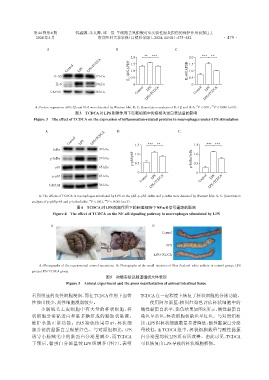
                   A:Photographs of the experimental animal operations. B:Photographs of the small intestine of New Zealand white rabbits in control group,LPS
                group,LPS+TCDCA group.
                                                  图5 动物实验及肠道组织大体表现
                                Figure 5  Animal experiment and the gross manifestation of animal intestinal tissue

                看到明显的炎性细胞浸润,而在TCDCA作用下血管                          TCDCA在一定程度上恢复了杯状细胞的分泌功能。
                性渗出较少,炎性细胞浸润较少。                                       使用阿尔新蓝⁃核固红染色评估杯状细胞中的
                    小肠绒毛上皮细胞中有大量的杯状细胞,杯                           酸性黏蛋白水平,染色结果如图8所示,酸性黏蛋白
                状细胞分泌黏蛋白和黏多糖组成的凝胶状黏液,                             染色呈蓝色,杯状细胞核染色呈红色。与对照组相
                维护小肠正常功能。PAS 染色结果显示,杯状细                           比,LPS组杯状细胞数量显著降低,酸性黏蛋白分泌

                胞分泌的黏蛋白呈现紫红色。与对照组相比,LPS                           量较低,而 TCDCA 组中,杯状细胞数量与酸性黏蛋
                诱导小肠绒毛中的黏蛋白分泌量减少,而 TCDCA                          白分泌量均较 LPS 组有所改善。由此可见,TCDCA
                干预后,黏蛋白分泌量较 LPS 组增多(图 7),表明                       可以恢复由LPS导致的杯状细胞损伤。
   36   37   38   39   40   41   42   43   44   45   46