Page 11 - 南京医科大学自然版
P. 11

第44卷第12期               程   丽,倪 曼,王 洁,等. 环磷酰胺对卵母细胞发育潜能影响的研究[J].
                 2024年12月                  南京医科大学学报(自然科学版),2024,44(12):1621-1628,1648                   ·1625 ·


                          A              Mock                  Ctrl                  4⁃HC
                               Cumulus cell expansion


                               PB1


                                                 100 μm               100 μm                100 μm

                               Blastocyst


                                                 100 μm               100 μm                100 μm
                                                                     *                         *
                          B            *              C                          D
                              200        *                  1.0        *              1.0        *
                                                                                      0.8
                                                            0.8
                             (μm)  150                     in vitro  0.6              0.6
                             Diameter  100               Maturation rate of GV  oocyte  0.4  Blastocyst rate  0.4

                               50
                                                                                      0.2
                                                            0.2
                               0                             0                         0
                                  Mock Ctrl 4⁃HC                Mock Ctrl 4⁃HC           Mock Ctrl 4⁃HC
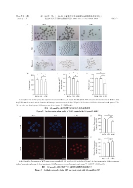
                   A:Compared with the Ctrl group,the expansion of cumulus cells in COC treated with 1.0 μmol/L 4⁃HC was poor,the extrusion rate of the first polar
                body(PB1)was decreased,and the formation of blastocyst was decreased(scale bar=100 μm). B:Cumulus cell diffusion diameter in each group. C:The
                                                          *
                PB1 extrusion rate of each group. D:Blastocyst rate of each group. P < 0.05(n=60).
                                           图2 1.0 μmol/L 4⁃HC作用下COC体外成熟培养结果
                                   Figure 2 In vitro maturationresults of COC treated with 1.0 μmol/L 4⁃HC

                   A           Mock                 Ctrl               4⁃HC           B      250      **  **
                                                                                         Relative fluorescence  intensity of GSH  150
                                                                                             200


                     GSH                                                                     100
                                                                                             50
                                                                                              0
                                                                                                 Mock Ctrl 4⁃HC
                                      150 μm               150 μm              150 μm  C             **
                                                                                            100         *
                                                                                         Relative fluorescence  intensity of DHE  60
                                                                                             80

                     DHE                                                                     40

                                                                                             20
                                                                                              0
                                      150 μm               150 μm              150 μm           Mock Ctrl 4⁃HC
                   A:ROS staining fluorescence of MⅡ stage oocytes treated with 1.0 μmol/L 4⁃HC(scale bar=150 μm). B:Semi⁃quantitative GSH fluorescence
                                                                                       **
                                                                                 *
                levels of oocytes in each group. C:Semi⁃quantitative DHE fluorescence levels of oocytes in each group. P < 0.05,P < 0.01(n=60).
                                        图3 1.0 μmol/L 4⁃HC作用下MⅡ期卵母细胞氧化应激水平
                                  Figure 3  Oxidative stress levels in MⅡoocytes treated with 1.0 μmol/L 4⁃HC
   6   7   8   9   10   11   12   13   14   15   16