


Page 89 - 南京医科大学自然版
P. 89
第45卷第10期        陈宣羽,朱 陈,林思颖,等. 吲哚菁绿近红外光成像示踪技术应用于达芬奇机器人胃癌根治术中D2淋巴
                 2025年10月            结清扫的安全性和有效性[J]. 南京医科大学学报(自然科学版),2025,45(10):1455-1466              ·1457 ·

                                                  表1   不同胃切除范围的注射点选择
                                      Table 1 Injection point selection for different gastrectomy ranges

                                                             Different gastrectomy ranges
                 Injection point
                                            Proximal gastrectomy                       Distal gastrectomy
                Lesser curvature  First branch of the right gastric artery  First branch of the right gastric artery
                              Gastric angle                                Gastric angle
                              Between the first and second gastric wall branches of the left Between the first and second gastric wall branches
                              gastric artery                               of the left gastric artery
                Greater curvature Terminal gastric branch of the right gastroepiploic artery  First gastric branch of the right gastroepiploic artery
                              First gastric branch of the left gastroepiploic artery  First gastric branch of the left gastroepiploic artery
                              Greater curvature at the fundus⁃body junction of the stomach  Midpoint between two injection sites


                                                              A                               B

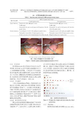
                                             A:Greater curvature injection. B:Lesser curvature injection.
                                               图1 机器人系统辅助下行ICG浆膜下注射
                                        Figure 1 Robotic system⁃assisted subplasma injection of ICG

                1.2.2  手术管理                                       后,对手术区域进行荧光成像,确保无任何残留的
                    使用配备 Firefly 荧光显影技术的达芬奇 Xi 手                  淋巴结。根据日本胃癌分类指南                [18] 对淋巴结站的
                术系统,通过 805 nm 近红外激发光源激活 ICG 分                     定义,对切除下的标本中分离出含淋巴结的软组
                子,实现精准淋巴结导航。手术过程中可以实现自                            织,标明所有淋巴结所属站别(图 2)。最后统一送
                然光和 NIR 荧光模式的随时切换,在近红外荧光模                         检病理科进行标准检验。
                式下可以实时、清晰地显示胃周淋巴结的轮廓及界
                                                                      NO.1
                限,从而有助于指导术者完整、彻底清扫规定范围                                     NO.3
                的淋巴结。所有手术均由同一团队完成。
                                                                                                          NO.4sb,
                1.2.2.1 手术操作
                                                                                                          NO.4d
                                                                              NO.5
                    手术方式根据肿瘤位置确定,包括近端胃、远
                端胃及全胃切除,切除后进行消化道重建(具体参                                                NO.6
                照日本胃癌治疗指南          [14] )。手术操作参照《机器人
                                                                                   NO.7
                胃癌手术中国专家共识(2021 版)》 和《腹腔镜胃
                                               [15]
                                                                       NO.8a
                癌手术操作指南(2023 版)》 ,D2 淋巴结清扫操作
                                        [16]
                依据江志伟教授提出“斯诺克路径” ,采用“由远                                                  NO.9,NO.11p
                                                [17]
                及近、由浅入深”顺序,遵循“整块切除”原则,保持                              NO.12a
                解剖层次的完整性。                                                    图2 远端胃切术后淋巴结分捡
                1.2.2.2 淋巴结清扫及分捡                                  Figure 2  Postoperative lymph node harvesting after distal
                    淋巴结清扫范围参照胃癌处理规约                 [18] 和《局部             gastrectomy
                进展期胃癌规范化淋巴结清扫范围中国专家共识                             1.2.3 观察指标
               (2022 版)》 。在 ICG 组中,完成所有淋巴结清扫                          一般资料:性别、年龄、体重指数(body mass index,
                         [19]

