Page 10 - 南京医科大学自然版
P. 10

第45卷第11期
               ·1540 ·                           南 京    医 科 大 学 学         报                        2025年11月


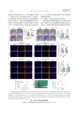
              敲降组则显著升高(图1G、H)。流式细胞术分析显                          低。以上结果表明,RBMS3在体外和体内均能抑制
              示:RBMS3 过表达可诱导 MDA⁃MB⁃231 和 SUM⁃                  乳腺癌细胞增殖。
              1315 细胞 G0/G1 期阻滞(图 2A~D)并显著提高细胞                  2.2  RBMS3上调乳腺癌细胞中p21的表达
              凋亡率(图2E~H)。裸鼠异种移植瘤模型结果显示:                              基于前期转录组测序数据的GO功能富集分析
              RBMS3 过表达组的肿瘤生长速度显著慢于对照组                          显示,RBMS3 表达与细胞增殖负向调控密切相关
             (图1I),且肿瘤体积(图1J)与质量(图1K)均显著降                      (图 3A),与 RBMS3 诱导细胞周期阻滞的实验结果
              A      NC      RBMS3   B                 C      SCR       Sh1       Sh2   D
                                                  NC                                                     SCR
                                       150  **    RBMS3                                          **      Sh1
                SUM⁃1315               Colony number  100  ***  SUM⁃1315                  Colony number  200  **  *** Sh2
                                                                                          150
                                                                                                        **
                                        50
                                                                                          100
                MDA⁃MB⁃231             SUM⁃1315           MDA⁃MB⁃231                       50 0  SUM⁃1315 MDA⁃MB⁃231
                                         0
                                           MDA⁃MB⁃231

              E                                                                          F
                               SUM⁃1315                            MDA⁃MB⁃231
                                                                                            (%)   NC
                     DAPI        EdU        Merge         DAPI        EdU        Merge       60   RBMS3
                                                                                            Rate of EdU positive cells  20
                NC                                    NC                                     40   **   ***


                RBMS3                                 RBMS3                                   0 SUM⁃1315 MDA⁃MB⁃231


              G                SUM⁃1315                            MDA⁃MB⁃231            H
                     DAPI        EdU        Merge         DAPI        EdU        Merge
                                                                                            (%)  SCR
                                                                                                 Sh1
                SCR                                   SCR                                    80  ***     ***
                                                                                            Rate of EdU positive cells  40
                                                                                                         ***
                                                                                                 Sh2
                                                                                                  ***
                                                                                             60
                Sh1                                   Sh1                                    20 0


                Sh2                                   Sh2                                      SUM⁃1315 MDA⁃MB⁃231


                          (mm 3 )  2 000  NC  **      NC                            (g)  1.5
                       I                           J                            K             **
                                 RBMS3
                          1 500
                          Tumor volumn  1 000 0  ***  RBMS3                         Tumor weight  0.5 0
                                                                                    1.0
                           500
                                   5
                                0
                                     10 15 21
                                   Time(d)                                                NC   RBMS3
                 A-D:Colony formation assay was performed to evaluate the colony⁃forming ability of SUM⁃1315 and MDA⁃MB⁃231 cells after overexpression(A,
              B)or knockdown of RBMS3(C,D)(n=3). E-H:EdU incorporation assay was used to detect cell proliferation after overexpression(E,F)or knockdown
              of RBMS3(G,H)(scale bar=200 μm)(n=3). I-K:In the SUM⁃1315 xenograft tumor model in nude mice,tumor volume was dynamically monitored in
              the RBMS3 overexpression group and the control group. Tumors were dissected,photographed,and weighed at the experimental endpoint(21 days after
                             **
              cell inoculation)(n=6). P < 0.01 and  *** P < 0.001.
                                                 图1  RBMS3抑制乳腺癌细胞增殖
                                     Figure 1  RBMS3 inhibited the proliferation of breast cancer cells
   5   6   7   8   9   10   11   12   13   14   15