
Page 18 - 南京医科大学自然版
P. 18
第45卷第11期
·1548 · 南 京 医 科 大 学 学 报 2025年11月
trifuges(Eppendorf,Germany);Cell incubator(Ther⁃ mixture was shaken vigorously for 15 s,followed by a
moFisher Scientific,USA);Chemiluminescence imager 3⁃minute rest,The sample was centrifuged at 12 000 g
(Tanon,Guangdong,China);Electrophoresis appara⁃ at 4 ℃ for 15 min. We transfered the upper layer of
tus,Thermal cycler(BIORAD,USA);Flow cytometer liquid to a new centrifuge tube,added 0.5 mL of isopro⁃
(Beckman Coulter,USA);Fluorescence microscope panol to precipitate the RNA follaued by a 10⁃minute
(Olympus,Japan);Microplate reader(Biotek,USA). rest,and then centrifuged the sample at 12 000 g at 4 ℃
1.2 Methods for 10 min. Subsequently,we discarded the supernatant
1.2.1 Lentiviral and plasmid transfections and then washed the RNA precipitate with 75% ethanol
shRNA and overexpression plasmids were de⁃ twice,and finally dissolved the RNA precipitate with
signed for genes including KIF11. They were transfect⁃ 20-50 μL of RNase⁃free water. SYBR Green premix
[18]
ed into cells according to the instructions . was used for qRT ⁃ PCR. The comparative threshold
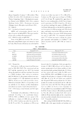
1.2.2 RNA Extraction and qRT⁃PCR cycle 2 - ΔΔCT method was used to evaluate the relative
Total cellular RNA was extracted. The cells were gene expression and normalize it to GAPDH [19] . The
fully lyzed using TRIzol reagent and allowed to stand primer sequences used for qRT ⁃ PCR are shown in
for 5 min. Then 0.2 mL of chloroform was added and the Table 1.
表1 引物序列表
Table 1 Primer sequences
Gene Forward(5′→3′) Rerverse(5′→3′)
KIF11 TTAATTTGGCAGAGCGGAAA CCATACGCAAAGATAGTGCAA
METTL3 GCTGACCATTCCAAGCTCTC ATTTCTTGGCTGGCTCCTTT
IGF2BP2 TTGCAGGAATTGACGCTGTA ACCCAAGGCGTTCAGATTTA
PROM1 TTTGCTGCTTGTGGAATAGAC ATAGGAAGGACTCGTTGCTGG
GAPDH GAAGGTGAAGGTCGGAGT GAAGATGGTGATGGGATTTC
1.2.3 Western blot detected after 2 h of incubation. Cells were spread at a
Total protein of cells was extracted and then sepa⁃ density of 1 000-2 000 cells per well for colony for⁃
rated by 10% SDS⁃PAGE. The concentrated gel was mation experiments in 6⁃well plates,and the cell colo⁃
80 V and the separated gel was 120 V for constant nies were fixed with paraformaldehyde after 10-14 days
voltage electrophoresis and then transferred the protein and then stained with crystal violet. For EdU experi⁃
to a PVDF membrane. After sealing the membrane ments,HCT116,DLD1,and SW480 cells were spread
with 5% BSA for 2 h,the primary antibody was incu⁃ evenly in 96⁃well plates at a density of 5 000 cells per
bated overnight at 4 ℃. The membrane was rinsed in well. After 24 h,the 96 ⁃ well plates were incubated
TBST for 10 min on day 2 and repeated three times, with 20 μmol/L EdU working solution for 2 h. Subse⁃
followed by incubation with the corresponding second⁃ quently,the medium was discarded,and the cells were
ary antibody for 2 h. Finally,an ECL chemilumines⁃ washed three times with PBS. Then 4% paraformalde⁃
cent solution was used to visualize the protein bands. hyde was added and fixed at room temperature for
1.2.4 CCK⁃8 assay,colony formation,and EdU exper⁃ 15 min,followed by permeabilization with 0.5% Triton
iments X⁃100 for 10 min to enhance cell membrane permeabil⁃
To determine cell proliferation,a CCK⁃8 test was ity. Next,the cells were incubated with click reaction
performed. According to the experimental design, buffer for 30 min away from light. and the cell prolifera⁃
HCT116,DLD1,or SW480 cells were spread in 96 ⁃ tion ratio was detected under a fluorescence microscope.
well plates at a density of 2 000 cells per well. After 1.2.5 Transwell experiments
24-36 h,CCK⁃8 solution was added for another 24, After adding 60 000-80 000 HCT116,DLD1,or
36,48,72,and 96 h. The absorbance at 450 nm was SW480 cells to the upper chamber and medium con⁃