
Page 14 - 南京医科大学自然版
P. 14
第45卷第2期
·154 · 南 京 医 科 大 学 学 报 2025年2月
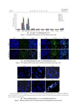
200 ABC⁃2
180 ABC⁃17
ABC⁃18
160 ABC⁃56
Fluorescence intensity 120 ssDNA pool
140
ABC⁃82
100
80
60
40
20
0
SKBR3 MCF⁃7 MDA⁃MB⁃231 MCF⁃10A SGC⁃7901 A549 SW480 A2780 HepG2 PANC⁃1
图5 DNA适配子与各种腺癌细胞的结合情况
Figure 5 The binding of DNA aptamers to various adenocarcinoma cells
DAPI ABC⁃18 Merge DAPI ABC⁃56 Merge
MCF⁃7 MCF⁃7
MDA⁃MB⁃231 MDA⁃MB⁃231
图6 激光共聚焦显微镜观察DNA适配子与乳腺癌细胞株的结合(×200)
Figure 6 DNA aptamers bind to breast cancer cell lines by confocal laser microscope(×200)
×1 000 ×1 000 ×1 000
A B C
×1 000 ×400 ×400
D E F
Analysiss of immunofluorescence staining. A⁃C:Breast cancer tissues with positive binding,showing Luminal A(A),Luminal B(B)and HER2(C)
subtype,respectively. D:Breast cancer tissues with negative binding(triple negative subtype). E,F:Breast fibroadenoma tissues(E)and normal breast
tissues(F)with low or negative binding.
图7 免疫荧光显微镜观察适配子ABC⁃56与乳腺来源的各种组织的结合
Figure 7 Binding of aptamer ABC⁃56 to tissues from breast by immunofluorescence microscope