


Page 111 - 南京医科大学自然版
P. 111
第45卷第4期          赵小静,张    丽,王 璐,等. 临床及内镜特征联合外周血炎症相关指标的评估在回肠末端
                  2025年4月          病变的病因诊断价值初探[J]. 南京医科大学学报(自然科学版),2025,45(4):544-550                   ·547 ·


                表 2  纳入研究的回肠末端病变患者内镜下表现与疾病谱                       两组在性别、吸烟史、腹部手术史、特殊药物使用史
                     分布                                           等其他人口统计学特征方面均无显著差异。
                Table 2  Endoscopic manifestations and disease spectrum
                                                                      将 22 例 CD 患者与 73 例非特异性回肠末端溃
                        distribution of patients with terminal ileum lesions
                                                                  疡患者按照性别和年龄进行 1∶1 匹配,比较两组患
                 Endoscopic findings                Number(n)
                                                                  者在初诊时的临床及内镜特征。结果发现,CD患者
                 Congestive erosion
                                                                  腹痛发生率显著高于非特异性回肠末端溃疡患者,
                   Ileitis of unknown etiology         521
                                                                  且更易表现为纵行溃疡及回盲瓣受累的内镜特征
                 Ulceration
                                                                 (P均<0.05)。而两组在发热、腹泻、体重下降、消化
                   Ileal ulceration with known etiology  020
                                                                  道出血、肠梗阻病史、肛周病变病史方面差异无统
                   CD                                  011
                   ITB                                 002        计学意义(表4)。
                   Lymphoma                            004        2.5  外周血炎症相关指标在CD与非特异性回肠末
                   UC                                  001        端溃疡患者的诊断价值
                   Adenocarcinoma                      285            比较两组患者初诊时的外周血炎症相关指标,
                   Ileal ulceration of indeterminate etiology  003
                                                                  发现 CD 组的 PLT、NLR、PLR 显著高于非特异性
                 Hyperplasia
                                                                  回肠末端溃疡组(P 均< 0.05),而 HB、ALB 则减低
                   Lymphoma                            051       (P 均< 0.05,表 5)。此外,ROC 曲线分析显示 PLT、
                   Benign tumor(including polyps)      057
                                                                  HB、ALB、NLR、PLR 等指标辅助回肠末端溃疡诊
                   Unknown etiology                    001
                                                                  断 为 CD 的 AUC 值 分 别 为 0.71、0.76、0.74、0.69、
                 Others *
                                                                                                             9
                   Macroglobulinemia                   956        0.75(图 2A、B),最佳临界值分别为 231.00×10 /L、
                 Total                                            130.21 g/L、37.30 g/L、1.86、168.39,灵敏度分别为
                   *Endoscopic findings:mucosal pallor,with gyriform changes and  63.6%、72.7%、95.5%、72.7%、86.4%,特异度分别
                villi disappearance.
                                                                  为77.3%、77.3%、59.1%、72.7%、63.6%(表6)。上述
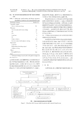
                出10例CD患者,本研究最终共纳入CD患者22例,非                        5 项指标联合辅助回肠末端溃疡诊断为 CD 的 AUC
                特异性回肠末端溃疡患者 73 例。两组患者初诊时                          值为0.83(图2C)。
                临床特征见表3。CD组年龄(33.9±12.1)岁,非特异
                                                                  3  讨 论
                性回肠末端溃疡组年龄(49.3±15.1)岁,CD组初诊年
                龄显著小于非特异性回肠末端溃疡组(P < 0.001)。                          本研究发现,绝大多数回肠末端病变内镜下仅


                                    Patients with terminal ileum
                                        lesions(n=956)
                                                                       Patients lost to follow⁃up(n=425)
                                                                       Ulceration(n=14)
                                                                       Congestive erosive(n=369)
                                                                       Proliferative lesions(n=42)

                             Diagnosis confirmed(n=93)  Diagnosis unconfirmed(n=438)


                     1. Polyp(n=46)
                     2. CD(n=20)              Ulceration(n=265)   Congestive erosive(n=158)  Proliferative lesions(n=15)
                     3. ITB(n=11)        (188 follow⁃up colonoscopics)  (93 follow⁃up colonoscopics)  (12 follow⁃up colonoscopics)
                     4. UC(n=4)
                     5. Benign tumor(n=5)
                     6. Lymphoma(n=5)         CD(n=10)     Disappearance of the lesions(n=116)
                     7. Adenocarcinoma(n=1)
                     8. Macroglobulinemia(n=1)
                                                          Without medication(n=73)

                                                图1   回肠末端病变患者随访结果流程图
                                 Figure 1  Flowchart of follow⁃up results for patients with terminal ileum lesions

