


Page 66 - 南京医科大学自然版
P. 66
第45卷第4期
               ·502 ·                            南 京    医 科 大 学 学         报                        2025年4月


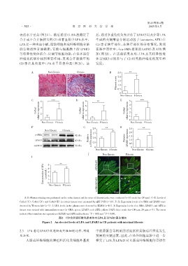
              表达水平更高(图 2H)。随后通过 ELISA 法测定了                      后,通过多重免疫荧光评估了LPAR3以及介导LPA
              合并或不合并肠狭窄的 CD 患者血浆中 LPA 水平,                       生成的关键酶自分泌运动因子(autotaxin,ATX)在
              LPA 是一种由血小板、脂肪细胞和成纤维细胞分泌                          CD 患者狭窄部位、非狭窄部位的分布情况,发现
              的生物活性甘油磷脂,它能与细胞膜上的 LPAR3                          在狭窄部位中,与α⁃SMA 相邻的 LAPR3 及 ATX 增
              等特异性受体结合,以调节细胞功能,在许多器官                            加(图 2J)。在这些结果表明,LPA 及其特异性受
              纤维化疾病中起到重要作用,发现合并肠狭窄的                             体 LPAR3 可能参与了 CD 相关肠纤维化的发生和
              CD 患者其血浆中 LPA 水平显著升高(图 2I)。最                      发展。


                      A          Non⁃fibrosis       Fibrosis             B     2.5    **
                                                                               2.0
                                                                              Fibrosis score  1.5

                                                                               1.0
                                                                               0.5
                                                                                0
                                         72 μm              72 μm
                                                                                 Non⁃fibrosis Fibrosis
                      C     6      ***             D     8       **             E     5 4     ***

                         Col1a1  relative expression  4 2  Col6a1  relative expression  6 4 2  Col6a3  relative expression  3 2


                            0                            0                            1 0
                             Non⁃fibrosis  Fibrosis       Non⁃fibrosis  Fibrosis       Non⁃fibrosis  Fibrosis
                      F                                 G                       H
                                                              2.0      **             2.5      **
                               Non⁃fibrosis  Fibrosis                                 2.0
                                                           Relative  α⁃SMA expression  1.0  Relative
                        α⁃SMA                     42 kDa      1.5                    expression  1.5
                        LPAR3                     40 kDa      0.5                     1.0
                        GAPDH                     36 kDa                             LPAR3  0.5
                                                               0                        0
                                                                Non⁃fibrosis  Fibrosis  Non⁃fibrosis Fibrosis
                                                           J         Non⁃fibrosis   Fibrosis
                      I          5      **

                              Concentration of LPA  (μmol/mL)  4 3 2 1  ATX/DAPI  100 μm  100 μm


                                 0
                                 Non⁃fibrosis  Fibrosis          α⁃SMA/LPAR3

                                                                                          20 μm
                                                                            20 μm
                 A,B:Masson staining was performed on the colon tissues and the score of fibrosis index were evaluated(n=10,scale bar=20 μm). C-E:Levels of
              Col1a1(C),Col6a1(D),and Col6a3(E)in colonic tissues were measured by qRT⁃PCR(n=10). F-H:Expression levels of α⁃SMA and LPAR3 were
              detected by Western blot(n=3). I:LPA levels in the plasma were detected by ELISA(n=10). J:Expression levels of α⁃SMA,LPAR3,and ATX in
              tissues were stained with immunofluorescence(α⁃SMA:green;LPAR3:red;ATX:yellow;DAPI:blue;scale bar=100 μm,20 μm,n=3). The arrow
                                                            **
              indicated that simultaneous expression of LPAR3 and ATX in fibroblasts. P < 0.01 and  *** P < 0.001.
                                        图2 CD合并肠纤维化患者体内LPA及LPAR3表达增加
                             Figure 2  An elevated levels of LPA and LPAR3 in CD patients with intestinal fibrosis


              2.3  LPA通过LPAR3促进肠成纤维细胞迁移、增殖                      中胶原蛋白等物质的过度沉积是肠道纤维化发生
              及活化                                               发展的关键因素,因此,在体外细胞实验中进一步
                  人肠成纤维细胞的增殖和活化及细胞外基质                           探究了 LPA 及 LPAR3 对人肠成纤维细胞的直接作

