


Page 67 - 南京医科大学自然版
P. 67
第45卷第4期         林俊杰,王 舒,赵小静,等. 双调蛋白通过上调LPA⁃LPAR3调控肠成纤维细胞活化促进克
                  2025年4月              罗恩病肠纤维化[J]. 南京医科大学学报(自然科学版),2025,45(4):498-508                   ·503 ·


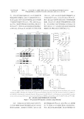
                用。首先,使用划痕实验来评估LPA对人肠成纤维                          (图3C~E)。此外,LPA促进人肠成纤维细胞产生更
                细胞迁移能力的影响,分别以不同浓度的LPA(0、5、                        多的胶原蛋白 Col1a1、Col6a1 和 Col6a3(图 3F~H)。
                10、20 μmol/L)处理人肠成纤维细胞,LPA显著提高                    随后,通过Ki67免疫荧光染色来特异性检测增殖期
                了划痕愈合的速度(图 3A、B)。同时,通过 Western                    细胞,评估 LPA 对人肠成纤维细胞增殖能力的影
                blot 评估了肠成纤维细胞活化标志物α⁃SMA 蛋白                       响。LPA 处理组中 Ki67 阳性细胞占比明显高于对
                的表达水平,发现 LPA 显著促进肠成纤维细胞中                          照组(图 3I、J),以上结果提示 LPA 促进人肠成纤维
                α⁃SMA 表达,在10 μmol/L 48 h条件下作用较为显著                 细胞的迁移、增殖、活化以及胶原蛋白产生。

                                                       LPA                                     ***
                                                                                 (μm 2 )  600 000  *  ***
                      A       Control    5 μmol/L     10 μmol/L  20 μmol/L   B    800 000
                                                                                 Migration area
                       0 h                                                        400 000
                                   72 μm       72 μm       72 μm        72 μm     200 000
                                                                                      0
                                                                                      Control
                       48 h                                                               5 μmol/L 10 μmol/L 20 μmol/L
                                   72 μm       72 μm       72 μm        72 μm
                                                                                               LPA
                      C                    LPA          D              ***          E            ***
                                Control 5 μmol/L 10 μmol/L 20 μmol/L  2.0  *  ***        2.0  ***

                         α⁃SMA                 42 kDa       α⁃SMA expression  1.5      α⁃SMA expression  1.5
                                                             1.0
                        GAPDH                  36 kDa        0.5                         1.0
                                                            Relative  0
                                  LPA 10 μmol/L                                          0.5
                              Control 24 h  48 h              Control                  Relative  0
                         α⁃SMA             42 kDa                 5 μmol/L 10 μmol/L 20 μmol/L  Control 24 h  48 h
                        GAPDH              36 kDa                      LPA
                      F        3       *          G     3      **          H      4      *
                              relative expression  1    1                         1
                            Col1a1  2                 Col6a1  relative expression  2  Col6a3  relative expression  3 2


                               0
                                 Control LPA(10μmol/L)  0  Control LPA(10μmol/L)  0  Control LPA(10μmol/L)
                                     Control           LPA
                      I                                                J     30      ***

                              DAPI/Ki67                                   Percentage of  Ki67 positive cells  20


                                                                             10

                                             20 μm            20 μm           0  Control  LPA(10μmol/L)
                   A,B:The migration ability of fibroblasts after treating with LPA was assessed by the wound healing assay(scale bar=72 μm,n=6). C-E:The
                expression of α⁃SMA in fibroblasts after treating with LPA was detected by Western blot(n=3). F-H:The expression of Col1a1(F),Col6a1(G),
                and Col6a3(H)of fibroblasts after treating with 10 μmol/L LPA was measured by qRT⁃PCR(n=3). I,J:Fibroblasts were stained with Ki67 immunofluo⁃
                                                                                        *
                                                                                              **
                                                                                                        ***
                rescence and the percentage of Ki67 positive cells was calculated(ki67:green;DAPI:blue;scale bar=20 μm,n=3). P < 0.05,P < 0.01,and P < 0.001.
                                            图3   LPA促进人肠成纤维细胞的迁移、活化和增殖
                                     Figure 3  LPA promoted HIF migration,activation,and proliferation
                    为进一步验证LPA是否通过LPAR3发挥作用,                       成纤维细胞迁移(图4A、B)、活化(图4C、D)、胶原蛋

                在以 LPA 刺激人肠成纤维细胞的同时加入 LPAR3                       白产生(图 4E~G)以及增殖(图 4H、I)的促进作用。
                抑制剂 Ki16425。Ki16425 显著抑制了 LPA 对人肠                 同时,通过转染 siRNA 敲减了肠成纤维细胞中的

