


Page 45 - 南京医科大学自然版
P. 45
第45卷第5期            路   寒,张雪飞,孙学伟,等. 两种小鼠模型在胆汁淤积性肝损伤实验中的应用[J].
                  2025年5月                     南京医科大学学报(自然科学版),2025,45(5):627-636                        ·631 ·


                           A              Control        Sham            BDL             ANIT


                              1 week


                              2 weeks


                           B              Control        Sham            BDL             ANIT


                               × 100

                             week
                             1

                               × 400


                           C              Control        Sham            BDL             ANIT


                               × 100
                             2 weeks


                               × 400


                           D
                                    5                                   5
                                  week  4                   BDL        2 weeks                   BDL
                                                                                                 ANIT
                                                            ANIT
                                  1  3                                  4 3       *     *
                                  Pathological score  2 1  *  *        Pathological score  2 1


                                    0                                   0
                                                                      Portal fibrosis
                                                                              Biliary proliferation
                                  Portal fibrosis Biliary proliferation  Inflammatory infiltration  Hepatic necrosis
                                  Inflammatory infiltration
                                                Hepatic necrosis


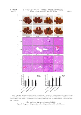
                   A:Gross morphological features of liver tissues across experimental groups. B,C:H&E staining of hepatic tissues at 1 week and 2 weeks intervals
                post⁃induction(black arrows:necrotic foci;yellow arrows:portal connective tissue hyperplasia;blue arrows:inflammatory cell infiltration;red arrows:
                ductular proliferation;×100,×400). D:Semiquantitative assessment of liver injury severity in the two experimental models. Compared to the ANIT
                    *
                group,P < 0.05(n=6).
                                          图2   BDL和ANIT模型肝脏病变的组织病理学对比分析
                            Figure 2  Comparative histopathological analysis of hepatic lesions in BDL and ANIT models

