


Page 53 - 南京医科大学自然版
P. 53
第45卷第6期     戈艳蕾,孙思怡,任泓沁,等. scRNA⁃seq和Bulk RNA⁃seq联合分析揭示MTRNR2L1介导COPD进展中
                  2025年6月              PANoptosis下调[J]. 南京医科大学学报(自然科学版),2025,45(6):786-797              ·791 ·


                                                表1 PANoptosis相关基因富集分析结果
                                      Table 1 GO enrichment analysis findings pertaining to PANoptosis
                    Data                                                              Gene ID
                    GO:0070269(pyroptosis)                                            TREM2
                    GO:1904019(epithelial cell apoptotic process)                    H19/BOK
                    GO:0034612(response to tumor necrosis factor)     CCL24/CALCA/GBP3/POSTN/ADAMTS12/VCAM1
                    GO:0006925(inflammatory cell apoptotic process)                   SLC7A11


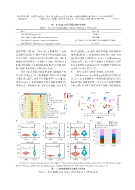
                组成可视化(图3A)。鉴于DEG 主要集中在与免疫                         胞、内皮细胞、上皮细胞、成纤维细胞、单核细胞和
                反应相关的过程中,故对各样本中免疫细胞组成进                            NK 细胞(图 3D)。结果发现 COPD 组中 CD8 T 细
                                                                                                           +
                                                            +
                行对比分析。结果发现COPD 患者肺组织中CD8 T                        胞占比显著低于对照组(P < 0.05),与 Bulk RNA⁃seq
                                                                                       +
                细胞的比例显著低于对照组(P < 0.05,图 3B);而 T                   分析的结果一致。CD8 T 细胞水平显著减少,表明
                细胞、NK 细胞、中性粒细胞、B 细胞、单核细胞的比                        存在异常免疫反应,提示 CD8 T 细胞参与的免疫反
                                                                                            +
                例在两组中未见明显异常(P均> 0.05)。                            应可能与 COPD 发生有关。
                    采用 UMAP 算法对两组样本中的细胞进行聚                        2.3  COPD患者肺组织MTRNR2L1表达增加
                类分析,共鉴定出 10 个细胞亚群(图3C),基于细胞                           Bulk RNA⁃seq 和scRNA⁃seq数据分析结果均显
                中基因表达模式,分析不同细胞类型中标志基因,                            示 COPD 患者肺部病变中免疫细胞浸润显著,表明
                采用 SingleR 对各类细胞亚群进行细胞类型注释,                       其免疫反应的潜在改变。为了证实上述测序数据
                共鉴定出 7 种细胞类型,分别是 B 细胞、CD8 T 细                     分析结果,对不同样本中 CD8 T 细胞、单核细胞和
                                                                                             +
                                                         +
                  A                                           Group     C            Control      COPD
                                            Group            4
                                                                Control                                     Count
                                            B cell              COPD
                                            Monocyte         2              10                                0
                                                                                                              1
                                            Macrophage/monocyte                                               2
                                                             0                                                3
                                            Myeloid dendritic cell           0                                4
                                            NK cell          -2           UMAP_2                              5
                                            T cell                                                            6
                                                                                                              7
                                            T cell CD8 +     -4                                               8
                                                                           -10
                                            cytotoxicity soore                                                9
                                            Cancer associaled fibroblast
                                            Neutrophil
                                                                             -15 -10 -5 0 5 10 -15 -10 -5 0 5 10
                                            Endothelial cell                              UMAP_1
                B                                                                   D
                         T cell CD8 +             T cell                NK cell           B cell       Fibrolasts
                                                               7.5                        CD8 T cell   Monocytes
                                                                                             +
                                                                                          Endothelial cells  NK cells
                  60                    40
                    Wilcoxon,P=0.029       Wilcoxon,P=0.066    5.0 Wilcoxon,P=0.330       Epithelial cells
                  40                                                                  100
                                        20                     2.5
                  20                                                                   75
                  0                      0                     0.0
                                                                                       50
                       COPD    Control        COPD   Control         COPD   Control
                                                                                       25
                         Neutrophil               B cell               Monocyte
                                                              250                      0
                 600
                                        40 Wilcoxon,P=0.052   200                            Control    COPD
                    Wilcoxon,P=0.730                                 Wilcoxon,P=0.26
                 400                    30                    150
                                        20                    100
                 200
                                        10                     50
                  0                      0                      0
                       COPD    Control        COPD   Control         COPD   Control
                   A:Heat map of immune cell composition in each sample. B:Violin chart of proportion of some immune cells in figure A. C:Cell annotation results
                for the COPD group and the control group. D:Proportions of different cell types in the COPD group(n=6)and the control group(n=15).
                                                图3   COPD患者肺免疫细胞浸润的改变
                                     Figure 3  Altered pulmonary immune cell infiltration in COPD patients

