
Page 13 - 《南京医科大学学报(自然科学版)》2025年第9期
P. 13
第45卷第9期 苏 宁,曹 颖,张淑平,等. PARP1在K192位点的乳酸化抑制卵巢癌细胞的迁移和增殖[J].
2025年9月 南京医科大学学报(自然科学版),2025,45(9):1219-1228,1241 ·1225 ·
A B
TIMELESS
WRN Zoom
PRKDC
PARP1 CASP7
XRCC1
XRCC5 CASP3
POLB XRCC6
PARG
C
PARP1:
PARP1 K192A:
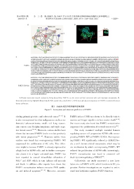
A:Protein interaction network analysis by String showed that PARP1 is at the center and had connection with each important oncoproteins. B:
Structural prediction by Alphafold showed that K192A caused only a small shift. C:K192 sites and adjacent sequences in PARP1 is conserved between
human and mouse.
图5 PARP1相互作用网络和结构预测
Figure 5 Interaction and structure prediction of PARP1
[19-21]
cluding primary prostate,and colorectal cancer . It PARP1 induced PAR was shown to be directly toxic to
[14- 29]
is also overexpressed in other malignancies,such as en⁃ neurons and trigger signals to induce neuron death .
dometrial adenocarcinoma,small ⁃ cell lung cancer, Our recent study also found that PARP1 overactivation
[14]
skin cancer,non⁃Hodgkin lymphoma,and triple⁃nega⁃ suppressed the proliferation of cervical cancer cells .
[22- 24]
tive breast cancer . Moreover,various studies have Our study examined multiple essential kinases
shown that increased PARP1 levels correlate positively regulating cancer cell progression A2780 cells overex⁃
with tumor progression [25- 26] . However,unlike these pressing PARP1⁃WT or PARP1⁃K192A. Overexpress⁃
studies,ours found that overexpressing PARP1 ⁃ WT ing PARP1⁃WT significantly reduced p⁃ERK1/2 lev⁃
suppressed the proliferation of OC cells. This differ⁃ els,a well ⁃ known critical oncoprotein,which may be
ence might be because PARP1 is already expressed at one mechanism by which overexpressing PARP1⁃WT
a high level in A2780 cells,and its further overexpres⁃ reduced the proliferation of A2780 cells. Interestingly,
sion places it in a hyper ⁃ activated state. PARP1 has PARP1 overactivation was found to promoted cell
[30]
been reported to caused intracellular exhaustion of death by downregulating p⁃ERK1/2 .
+
NAD and ATP,which in turn induces cell necrosis Collectively,our study uncovered a new lacty⁃
and death. In addition,other reports have shown that lation site on PARP1 in OC,which functions in OC pro⁃
PARP1 overactivation induces excessive PAR synthe⁃ gression. Further investigations are required to under⁃
[27- 28]
sis and induced apoptosis . Moreover,excessive stand how lactylation regulates PARP1 function.