
Page 15 - 《南京医科大学学报(自然科学版)》2025年第9期
P. 15
第45卷第9期 苏 宁,曹 颖,张淑平,等. PARP1在K192位点的乳酸化抑制卵巢癌细胞的迁移和增殖[J].
2025年9月 南京医科大学学报(自然科学版),2025,45(9):1219-1228,1241 ·1227 ·
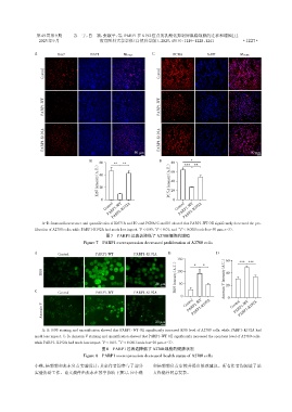
A Ki67 DAPI Merge C PCNA DAPI Merge
Control Control
PARP1⁃WT PARP1⁃WT
PARP1⁃K192A PARP1⁃K192A
50 μm 50 μm
B 60 ** ** D 80 *** * **
(A.U.) 40 (A.U.) 60
lntensity 20 PCNA lntensity 40
Ki67 20
0 0
Control PARP1⁃K192A Control PARP1⁃K192A
PARP1⁃WT
PARP1⁃WT
A-D:Immunofluorescence and quantification of Ki67(A and B),and PCNA(C and D)showed that PARP1⁃WT OE significantly decreased the pro⁃
**
*
liferation of A2780 cells,while PARP1⁃K192A had much less impact. P < 0.05,P < 0.01,and *** P < 0.001(scale bar=50 μm,n=3).
图7 PARP1过表达降低了A2780细胞的增殖
Figure 7 PARP1 overexpression decreased proliferation of A2780 cells
A B D
Control PARP1⁃WT PARP1⁃K192A
150 60 *** ***
(A.U.) * * (A.U.)
ROS 100 40
ROS lntensity
20 μm 50 Annexin V lntensity 20
C Control PARP1⁃WT PARP1⁃K192A
0 Control 0
PARP1⁃K192A
PARP1⁃WT
Annexin V PARP1⁃WT Control PARP1⁃K192A
20 μm
A,B:ROS staining and quantification showed that PARP1⁃WT OE significantly increased ROS level of A2780 cells,while PARP1⁃K192A had
much less impact. C,D:Annexin V staining and quantification showed that PARP1⁃WT OE significantly increased the apoptosis level of A2780 cells,
*
***
while PARP1⁃ K192A had much less impact. P < 0.05, P < 0.001(scale bar=20 μm,n=3).
图8 PARP1过表达降低了A2780细胞的健康状况
Figure 8 PARP1 overexpression decreased health status of A2780 cells
小燕、杨莉莉和张东负责实验设计;其余作者均参与了部分 和杨莉莉负责审校并提出修改建议。所有作者均阅读了论
实验协助工作。论文稿件由张东在苏宁协助下撰写,应小燕 文终稿并同意发表。