


Page 134 - 南京医科大学自然版
P. 134
第46卷第10期
               ·1564 ·                           南 京    医 科 大 学 学         报                        2026年10月


                                              A                           B                           C


                                              D                           E                           F


                                              G                           H                            I
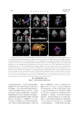
                 A:Parasternal short⁃axis view on echocardiography shows the main pulmonary artery and its left and right branches. The main pulmonary artery is
              dilated,and the diameter of the right pulmonary artery is smaller than that of the left pulmonary artery. MPA:main pulmonary artery;RPA:right pulmo⁃
              nary artery;LPA:left pulmonary artery. B:Subxiphoid biatrial view on echocardiography presents an enlarged right atrium. The right inferior pulmonary
              vein drains into the inferior vena cava. RA:right atrium;LA:left atrium;IVC:inferior vena cava;RIPV:right inferior pulmonary vein. C:Non⁃standard
              subxiphoid view on echocardiography shows the right pulmonary veins connecting to the inferior vena cava. RPV:right pulmonary veins. D:Non⁃stan⁃
              dard subxiphoid biatrial view on echocardiography shows the right inferior pulmonary vein directly entering the lower segment of the right atrium. E:
              CDFI shows persistent mosaic venous flow at the anastomotic site. CW Doppler reveals a bimodal laminar flow pattern with elevated flow velocity,and
              the maximum velocity is 156 cm/s. F,G:Subxiphoid view on echocardiography shows systemic⁃pulmonary collateral circulation arising from the abdominal
              aorta. AA:abdominal aorta;SPC:systemic⁃pulmonary collateral circulation. H:Three⁃dimensional reconstruction of CTA demonstrates that the right
              inferior pulmonary vein connects to the inferior vena cava,while the right superior pulmonary vein,left superior pulmonary vein and left inferior pulmo⁃
              nary vein drain into the left atrium. RSPV:right superior pulmonary vein;LSPV:left superior pulmonary vein;LIPV:left inferior pulmonary vein. I:CTA
              shows the right pulmonary veins converging into a common trunk and connecting to the inferior vena cava.
                                                   图1 典型病例的超声心动图
                                              Figure 1 Echocardiogram of typical cases


              房,此即“满足性偏误”。另外本组3例CDFI明确下                         晰显示下腔静脉走行。事实上,异常连接的“弯刀
              腔静脉处异常血流,因检查者经验不足被忽略,最                            静脉”常走行于右心房后方,引流点位于膈肌附近
              终导致漏诊。甚至 2 例仅诊断原发性肺动脉高压,                          的下腔静脉或右心房下部,该区域在常规心尖四腔
              本组低年资医师漏诊率高达 76%(19/25)。②标准                       心、胸骨旁长轴等切面中属于盲区或显示不清区
              化扫查缺失:这是最主要的漏诊原因。超声医师未                            域。检查者未主动采用剑突下切面进行针对性探
              能将肺静脉系统,特别是右肺静脉的全程追踪纳入                            查肺静脉及上、下腔静脉。这也与曹睿等                   [12] 漏诊的
              常规检查思维,漏诊病例的报告多数没有提示肺静                            情况类似。③疾病的不典型征象:由于检查者固定
              脉的走行,仅3例描述了4支肺静脉“汇入左心房”,                          思维,认为SS都具备相对典型的临床与超声特征组
              但未提供右下肺静脉入左心房的直接切面证据。                             合,即右心房室扩大,心脏右移/中位心,右肺动脉偏
              另外,回顾性分析漏诊病例的图像时,13 例没有清                          窄与肺动脉高压等。一旦检查时发现心脏位置未

